
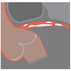
人工知能(AI*)による石灰化組織の分布や厚みの自動検出・自動測定
石灰化病変は、手技成功率の低下とステント拡張不良に起因しており、臨床予後の低下要因のひとつです1。
冠動脈での石灰化病変の有病率と重症度は上昇傾向にあり、患者の25%に中等度から重度の石灰化病変が認められます2, 3。
- 石灰化病変によるステント拡張不良は、ステント血栓症やステント内再狭窄の主要な予測因子とされています1。
- 良好なステント拡張は、PCI施行時の主要心血管イベントの発生率を軽減させることが示唆されています4。
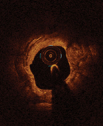
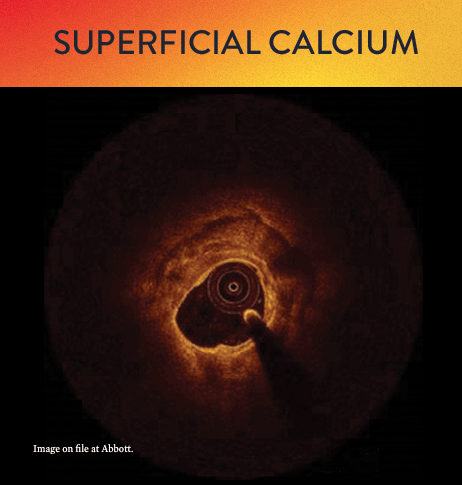
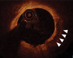
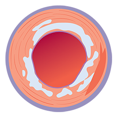
左:ほぼ360度の表在性石灰化
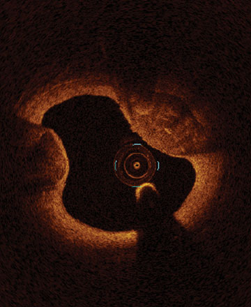
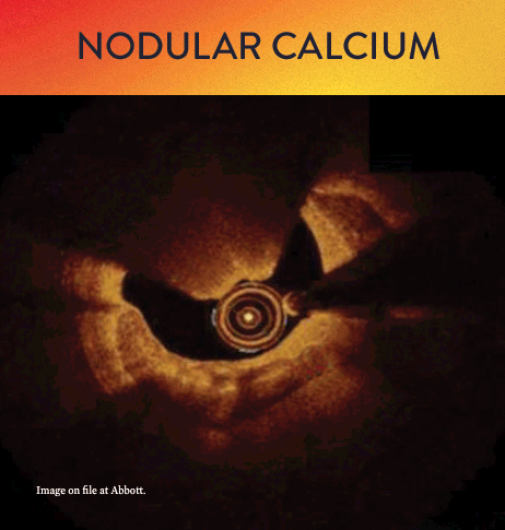
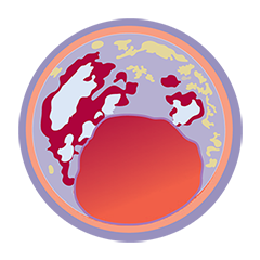
中:12時~3時方向と8時~10時方向の石灰化結節
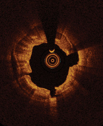
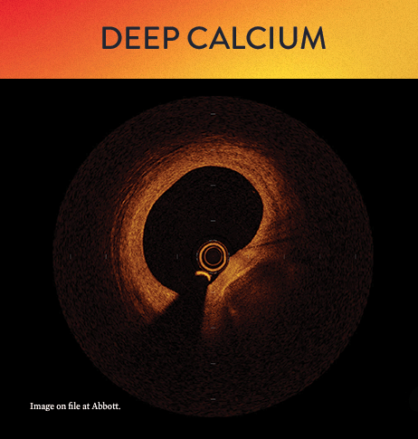
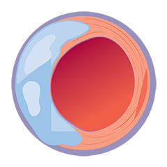
右:360度の表在性石灰化で3時、6時、9時方向のフラクチャー
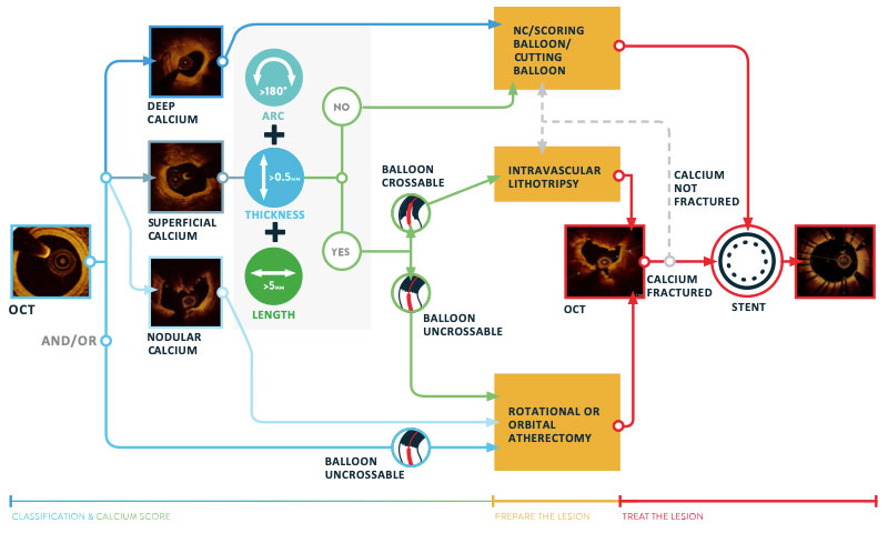
OCTによる石灰化の診断方法と治療方針
OCTイメージングは、至適な病変前処置(Lesion Preparaion)戦略に必要な石灰化病変の組織性状を視覚的かつ定量的に提供します1。
OCTワークフローの MLD-MAX は、迅速で一貫した手技と良好な予後のためのアルゴリズムです。このワークフローの最初のステップでは、Morphology(M:組織性状)を評価することにより、治療領域のプラーク性状を把握し、ステント留置前の至適な前処置方法を検討します。石灰化病変では、石灰化プラークの種類とその重症度を判断する上で役立ちます。

LightLab Clinical Initiative5 では、医師がOCTを用いて病変組織性状と重症度を評価すると、約3分の1の病変で前処置方針が変更されました。主に前処置方針の変更が確認された組織性状は、石灰化病変でした。
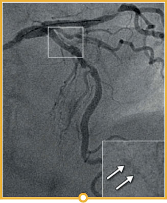
LIGHTLAB STUDYの結果では、PCI施行前の血管造影評価で計画されていた治療アプローチは(左画像)、術前のOCT評価の実施により変更されました(右画像)。
影響のあった領域は以下の通りです。
- 血管造影では、石灰化病変の程度が低く評価されていた。
- 前処置(Vessel preparation)戦略が、コンプライアントバルーンの使用ではなく、ノンコンプライアントバルーンと回転式アテレクトミーの使用に変更された。
- 血管造影評価で計画されていたステントサイズが、OCT評価の実施により6 mmサイズアップされた。
Morphology
(形態):
Length
(長さ):
Diameter
(直径):
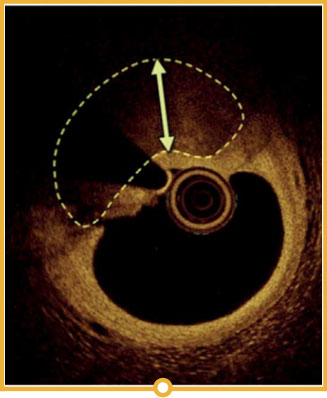
LAD中央部の石灰化病変(タイプB)
血行動態に影響あり
30 mm
遠位部3.0mm、近位部3.5mm
計画されていた血管前処置:コンプライアントバルーン
計画されていた治療:3.0mm x 32mmステント
Morphology
(形態):
Length
(長さ):
Diameter
(直径):
LAD中央部の石灰化病変(タイプB)
38 mm
遠位EEL 2.82mm、近位EEL 3.75mm
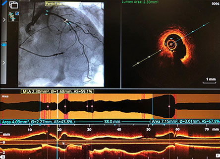
最小内腔面積: 2.3 mm²
OCTで計画された血管前処置:ノンコンプライアントバルーン、回転式アテレクトミー
計画された治療: 3.0 mm x 38 mm ステント
OCTによる石灰化評価
血管内イメージングを使用すると、血管造影による石灰化分類(軽度、中等度、重度)と比べ、詳細な石灰化病変の組織性状評価と定量化が可能となります。
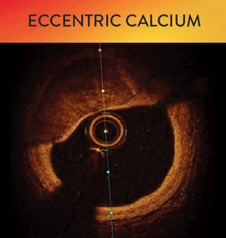
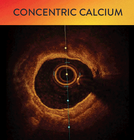
OCTは、石灰化を深在性、表在性、結節性に分類できるだけでなく、偏心性、同心性といった評価も可能です。MLD-MAXワークフローの最初のステップでは、石灰化病変の組織性状評価を行います。石灰化病変の組織性状により治療法が異なるため、M(Morphology:組織性状)は、至適な治療戦略を検討する上で重要な要素となります1。
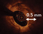
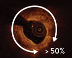
OCTによる石灰化スコアを基準としたRule of 5s
OCTでの石灰化スコア評価アルゴリズムを用いて石灰化の厚みを計測することで、ステント留置前に前処置が必要な石灰化病変の特定に役立ちます6。このアルゴリズムでは以下の点が、アテレクトミーデバイスなどの必要性を予測するのに用いられる重要なパラメータとなります。
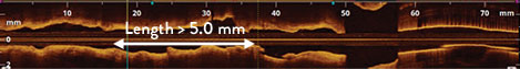
このアルゴリズムで用いられるパラメータでは、石灰化の厚さ、石灰化の角度、(長軸上の)石灰化の長さで評価します。
- 厚さ:0.5mm超
- 角度:血管アークの50%超
- 長さ:5mm超
ステント拡張不良のリスクがある病変は、石灰化スコアが46
OCTによる石灰化スコア
1 石灰化の最大角度
90度以下 0ポイント
90度超 < 角度 ≤ 180度以下 1ポイント
180度超 2 ポイント
2 石灰化の最大厚さ(mm)
0.5 mm以下 0ポイント
0.5 mm超 1 ポイント
3 石灰化の連続した長さ(mm)
5.0 mm以下 0ポイント
5.0 mm超 1 ポイント
総スコア = 0~4 ポイント基準ルール(Rule of 5s)
❶厚さ0.5mm
❷ 角度50%
❸長さ5.0mm
石灰化の厚さの評価は、ステント拡張の予測で重要です7。IVUSでは、超音波が石灰化病変ではね返り暗い影となります。OCTは、近赤外線が石灰化病変を透過するため、石灰化病変の厚さを評価することができます7。
OCTによる石灰化病変の診断・治療例
- MLD-MAXワークフローでPCIを開始します。
- 組織性状(M)を評価してプラークの種類を判定します。
- 石灰化プラークに対しては、OCTガイドによる石灰化病変アルゴリズムでの治療を行います1。このアルゴリズムによって、石灰化病変の評価から病変の前処置や至適ステント拡張を得るための情報が提供されます。
- OCTガイドを用いることで、石灰化の特性と血管の組織性状を正確に把握することができるため、至適な病変の前処置方針を確立しやすくなり、PCIの手技成功と、再治療の減少が期待されます。
石灰化病変に対するOCTイメージング
OCTは、石灰化の検出・位置特定・定量化に適した画像診断法のひとつです8。
冠動脈造影法、冠動脈コンピュータ断層撮影法(CT)、血管内超音波法(IVUS)、高周波法(RF)、血管内超音波法-バーチャルヒストロジー(IVUS-VH)および光干渉断層撮影法(OCT)はいずれも、石灰化を検出し、その位置を特定して定量化することが可能です。
Gary Mintz医師は、石灰化と冠動脈硬化症との関連性を対象とした20年に及ぶ血管内イメージング研究を通して、その診断精度は大きく異なると示唆しています8。
| 冠動脈造影法 | CT | IVUS | RF-IVUS (IVUS-VH) | OCT | |
|---|---|---|---|---|---|
| 画像診断法 | 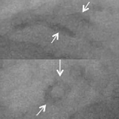 |  | 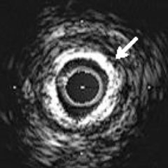 | 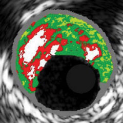 | 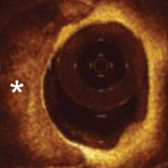 |
| 冠動脈石灰化の検出 | + | +++ | +++ | +++ | ++++ |
| 冠動脈石灰化の位置特定 | + | +++ | +++ | +++ | ++++ |
| 冠動脈石灰化の定量化 | + | +++ | ++ | +++ | ++++ |
 |  |  |  |  |
PCIの手技成功と再治療率の低下は、最終的なステント拡張と密接な関連があります1。血管造影では、治療方針に影響を与えうる病変組織性状の重症度を低く評価する可能性があるため、石灰化病変が正確に評価されない場合もあります5。
OCTイメージングとMLD-MAXワークフローの併用により、適切な治療アプローチの決定と、至適なステント拡張を支援します。LIGHTLAB STUDYでは、MLD-MAXワークフローを用いることで、平均80%の最小ステント拡張が得られました。

*Ultreon™機能の一部
参考文献
- Shlofmitz, E., Sosa, F. A., Ali, Z. A., Waksman, R., Jeremias, A., & Shlofmitz, R. (2019). OCT-Guided Treatment of Calcified Coronary Artery Disease: Breaking the Barrier to Stent Expansion. Current Cardiovascular Imaging Reports, 12(8), 32.
- Dean Kereiakes, Disrupt CAD III: Lithotripsy for Vessel Preparation in Calcified Coronary Arteries Prior to Stenting, CLD. Dec 29, 2020.
- Madhavan MV, Tarigopula M, Mintz GS, Maehara A, Stone GW, Généreux P. Coronary artery calcification: pathogenesis and prognostic implications. J Am Coll Cardiol. 2014 May 06;63(17):1703-14.
- Räber L, et al. Clinical use of intracoronary imaging. Part 1: guidance and optimization of coronary interventions. An expert consensus document of the European Association of Percutaneous Cardiovascular Interventions. Eur Heart J. 2018;39(35):3281-3300.
- Croce, K. et al: Optical Coherence Tomography Influences Procedure and Vessel Preparation Decisions During Percutaneous Coronary Intervention– Insights from the LightLab Initiative. TCTConnect2020 Presentation.
- Fujino, A et al. A new optical coherence tomography-based calcium scoring system to predict stent underexpansion. EuroIntervention. 2018 Apr 6;13(18):e2182-e2189. doi: 10.4244/EIJ-D-17-00962.
- Wang, X et al. In vivo calcium detection by comparing optical coherence tomography, intravascular ultrasound, and angiography. JACC Cardiovasc Imaging. 2017 Aug;10(8):869-879. doi: 10.1016/j.jcmg.2017.05.014.
- Mintz,G. Intravascular Imaging of Coronary Calcification and its Clinical Implications. JACC Cardiovascular Imaging, 2015. http://dx.doi.org/10.1016/j.jcmg.2015.02.003
販売名:SJM FD-OCT Integratedイメージングシステム
医療機器承認番号:22700BZX00153000
分類:管理医療機器
販売名:SJM FD-OCT イメージングシステム
医療機器承認番号:22300BZX00306000
分類:管理医療機器
販売名:SJM OCT イメージングカテーテル
医療機器承認番号:22300BZX00307000
分類:高度管理医療機器
販売名:ドラゴンフライ オプスター OCT イメージングカテーテル
医療機器承認番号:30100BZX00237000
分類:高度管理医療機器
MAT-2112780 v4.0