One Software. The Best of All Worlds.
Powered by AI, Ultreon™ 2.0 Software instantly provides you with actionable OCT and angiographic insights, enabling PCI procedural efficiencies and optimal outcomes.1-5
Ultreon™ 2.0 Software boasts:1,6
- The power of artificial intelligence (AI) from Ultreon™ 1.0 Software
- Similar user interface from AptiVue™ Software
- Dynamic Angio that enhances angiography
- OCT and Physiology capabilities
Be one of the first to experience Ultreon™ 2.0 Software
Ultreon™ 2.0 Software partners with you through every step of your PCI journey to enable easy, fast and accurate clinical decisions.
Customize the user profile for the best workflow for you1
Select the workflow mode, pullback trigger type, calcium threshold, expansion threshold and more before the procedure starts. You can always change these settings during a case too, if needed.
Plan the procedure with actionable insights powered by AI1
Let the power of AI help identify calcium burden and External Elastic Lamina (EEL) to guide the decision on the best vessel prep and stent strategy. AI-processed information is readily available as soon as the pullback loads.
Calcium is associated with decreased procedural success and stent underexpansion, resulting in adverse clinical outcomes.7
Place the stent where you planned it with the aid of Dynamic Angio1
The Dynamic Angio view enables users to see a live angio feed next to the co-registration view with their stent plan. Zoom in on both up to 600% to guide the most precise stent delivery and deployment possible.
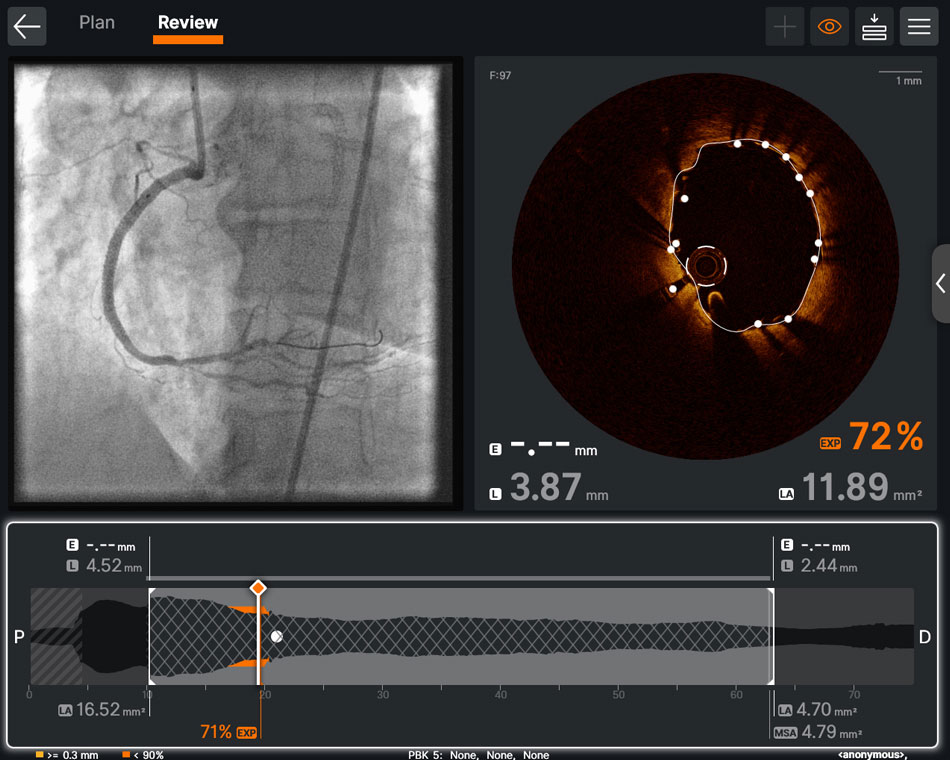
Confirm your work in seconds with automated expansion calculation1
The Review screen displays the expansion percentage for your post-PCI OCT run. Expansion lower than set threshold is highlighted in orange and malapposition larger than set threshold is highlighted in yellow. Users can quickly decide if additional optimization is needed.
Stent expansion impacts outcomes. Achieving optimal stent expansion is proven to reduce rates of adverse cardiac events. Stent underexpansion is an indicator of adverse events, such as stent thrombosis and restenosis.8
Analyze complex PCI cases with 3D OCT1
Access the new and powerful 3D Bifurcation View in
Ultreon™ 2.0 Software for an immersive view.
The 3D View allows physicians to visualize the vessel in a comprehensive way and guide their clinical actions with clarity.
Stent implantation in complex bifurcation lesions involving both the main vessel and side branch is associated with greater risk of procedural complications and worse clinical outcomes.9 In the OCTOBER trial, OCT-guided PCI reduced MACE by 28% in complex bifurcation lesions compared to angiography.9
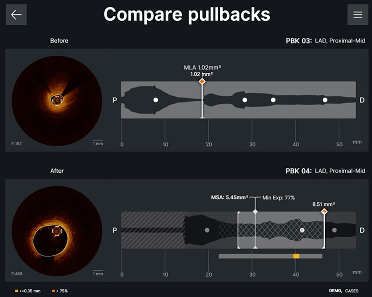
See the before and after with one click1
Compare any two pullbacks with the same vessel location and label to visually see the change of the vessel. Export the Compare pullbacks screen for easy sharing.
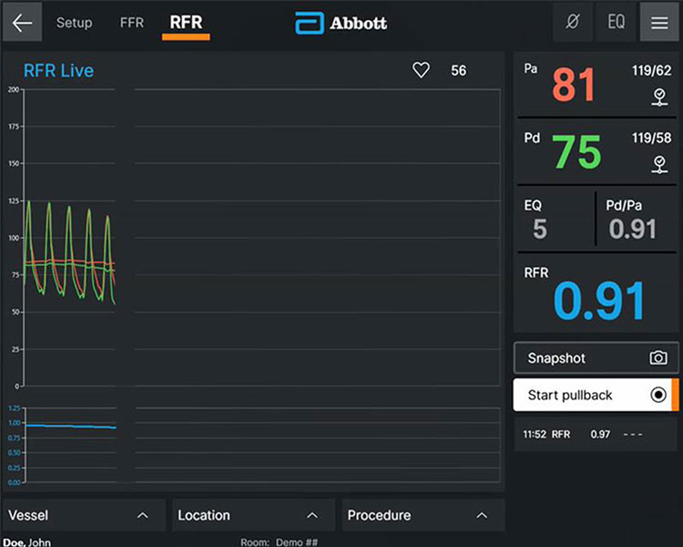
One software for both OCT and Physiology1
Ultreon™ Software offers a comprehensive diagnostic solution to measure physiological indices at stress (FFR) or rest (RFR).
With the redesigned user-friendly interface in
Ultreon™ 2.0 Software, you can:
- Easily start imaging or physiology for a selected case
- Easily toggle between FFR and RFR mode
- Appreciate large physiology views for rapid waveform assessment and drift checking
From Our Users Around the Globe


References
1. Ultreon™ 2.0 Software Instructions for Use (IFU). Refer to IFU for additional information.
2. Ali, Z. A., et al. (2016). Optical coherence tomography compared with intravascular ultrasound and with angiography to guide coronary stent implantation (ILUMIEN III: OPTIMIZE PCI): a randomised controlled trial. The Lancet, 388(10060), 2618–2628. https://doi.org/10.1016/s0140-6736(16)31922-5.
3. Prati F, et al. (2015). Clinical Impact of OCT Findings During PCI. Jacc-cardiovascular Imaging, 8(11), 1297–1305. https://doi.org/10.1016/j.jcmg.2015.08.013.
4. Rauch J, et al. (2021). TCT-276 A Standardized Optical Coherence Tomography Workflow Improves Procedural Efficiency and Safety During Percutaneous Coronary Intervention: Insights From the LightLab Initiative. Journal of the American College of Cardiology, 78(19), B113. https://doi.org/10.1016/j.jacc.2021.09.1129.
5. Lee J. M., et al. (2023). Intravascular Imaging–Guided or Angiography-Guided Complex PCI. The New England Journal of Medicine. https://doi.org/10.1056/nejmoa2216607.
6. AptiVue™ E.5.2 Software Instructions for Use (IFU). Refer to IFU for additional information.
7. Shlofmitz, E., Sosa, F. A., Ali, Z. A., Waksman, R., Jeremias, A., & Shlofmitz, R. (2019). OCT-Guided Treatment of Calcified Coronary Artery Disease: Breaking the Barrier to Stent Expansion. Current Cardiovascular Imaging Reports, 12(8), 32.
8. Räber L, et al. Clinical use of intracoronary imaging. Part 1: guidance and optimization of coronary interventions. An expert consensus document of the European Association of Percutaneous Cardiovascular Interventions. Eur Heart J. 2018;39(35):3281-3300.
9. N.R. Holm et al., OCT or Angiography Guidance for PCI in Complex Bifurcation Lesions, NEJM, DOI: 10.1056/NEJMoa2307770 (OCTOBER).
MAT-2303965 v1.0