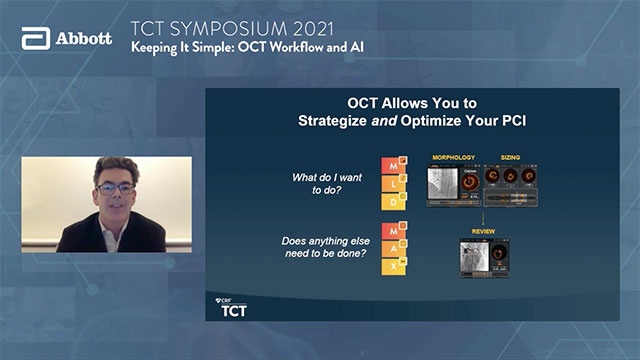
Streamline PCI Workflow with Accurate Stent Placement
Ultreon™ 1.0 Software guides physicians through percutaneous coronary intervention (PCI) step-by-step following MLD MAX workflow and provides insights on morphology, vessel sizing, stent placement and post-stent optimization for more accurate decision-making.1
Pre-PCI Guidance with Ultreon™ 1.0 Software
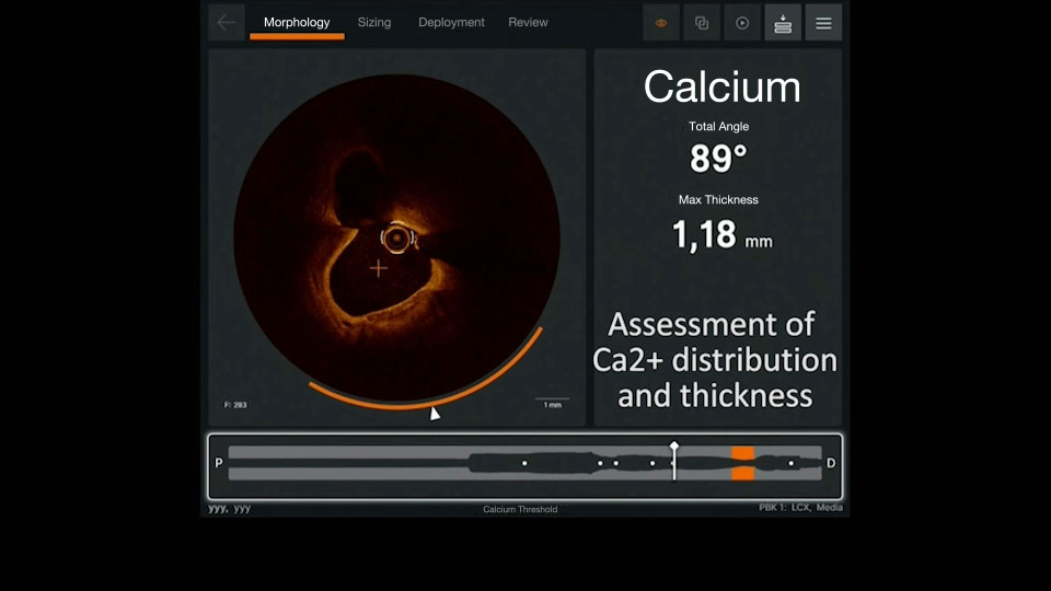
Morphology: Automatic calcium detection2
1 in 4 patients undergoing PCI, have moderate to severe calcium.3 If left undiagnosed and untreated, it adversely impacts PCI outcomes.4 Calcified lesions limit stent expansion, which is a major predictor of stent failure, such as stent thrombosis and restenosis.5
Ultreon Software displays calcification on the morphology screen:
- An orange arc around the cross-sectional view indicates the presence of calcium
- Arc is displayed when calcium angle is at or above 60 degrees of circumferential calcium — Learn more about how to identify calcium with OCT
- The white triangle, marked by the red circle, in the cross-sectional view indicates maximum calcium thickness [in the current frame]
- Total angle and max thickness values are displayed in real time throughout pullback
- Co-registration allows the user to visualize the calcium on the angio still frame
When calcification assessment is complete, a user can click on the Sizing tab to proceed to the next step of stent sizing evaluation.
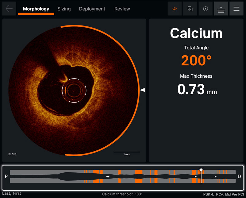
Morphology screen displaying angle and max thickness of calcification
Morphology screen displaying angle and max thickness of calcification
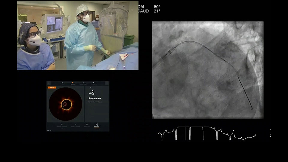
Watch how physicians use Ultreon™ 1.0 Software to assess calcific plaque and plan treatment approach
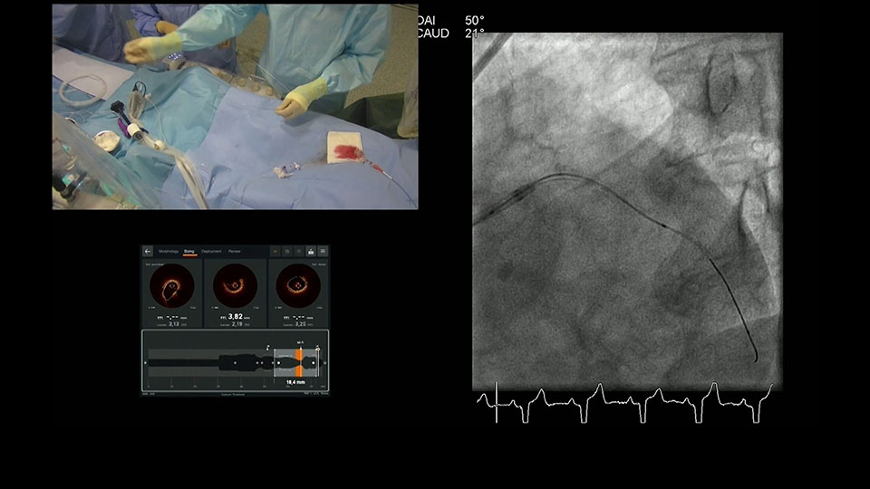
Sizing: Automatic detection of vessel diameter via EEL or Lumen2
Ultreon™ 1.0 Software uses artificial intelligence (AI) to automatically detect external elastic lamina (EEL) and lumen to help identify landing zones for accurate stent placement.1,6
- Length: Angiography co-registration facilitates identification of accurate length
- Diameter: External elastic lamina (EEL) and lumen help identify landing zones in healthy tissue for stent placement6
- Lumen shown as a solid white or red contour line
- EEL shown as the dotted white line in the cross-sectional image and lumen profile
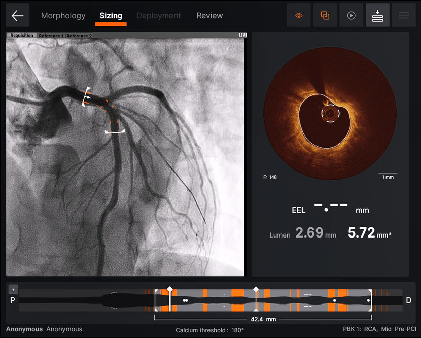
Ultreon™ 1.0 Software sizing screen displays sizing for vessel length and diameter
Ultreon™ 1.0 Software sizing screen displays sizing for vessel length and diameter
Sizing screen displays a four panel view:2
- Current selected frame
- Proximal reference frame
- Distal reference frame
- Maximized lumen profile – user can maximize the lumen profile by clicking on the icon at top left corner of lumen profile view.
Once sizing assessment is complete, a user can move to the next step by clicking the Deployment tab.
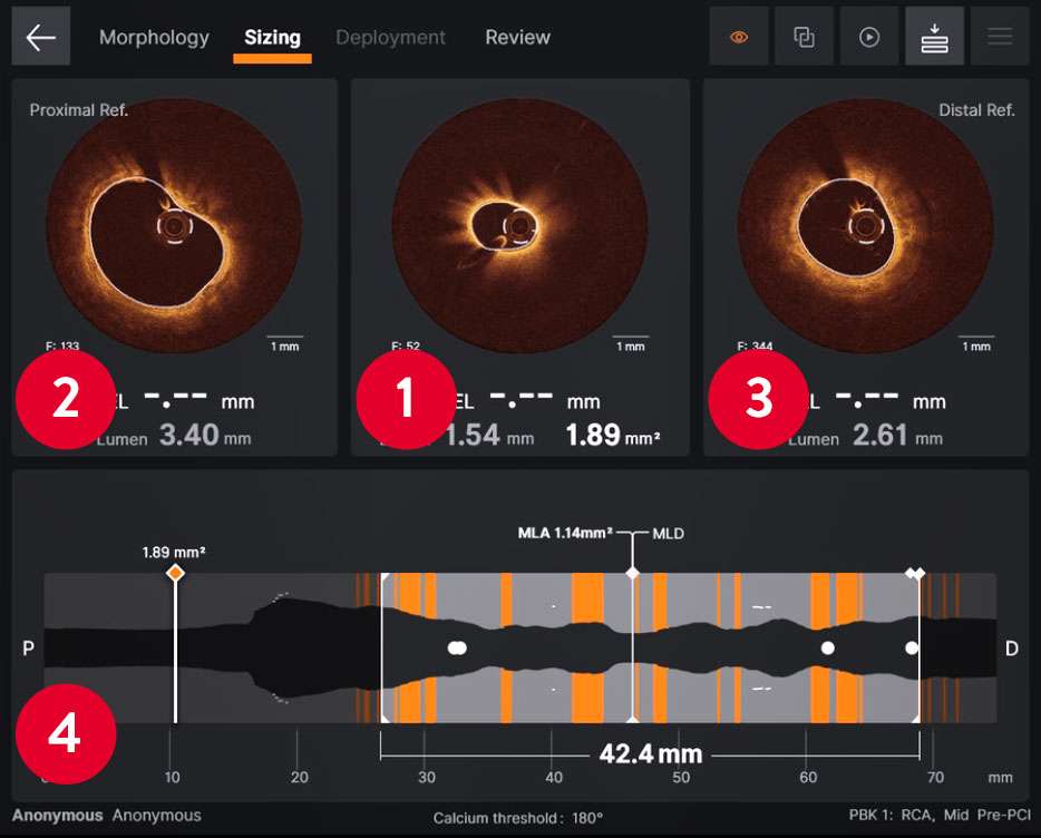
Ultreon™ 1.0 Software displays a four‑panel view sizing screen
Ultreon™ 1.0 Software displays a four‑panel view sizing screen
Deployment: Facilitation of precise stent deployment2
Ultreon™ 1.0 Software facilitates accurate stent deployment through co-registered and live angio views side-by-side.
- Users can select a full-screen co-registered view if desired
Once the stent deployment is complete, a user can move to the next step by clicking the Review tab for post-PCI optimization.
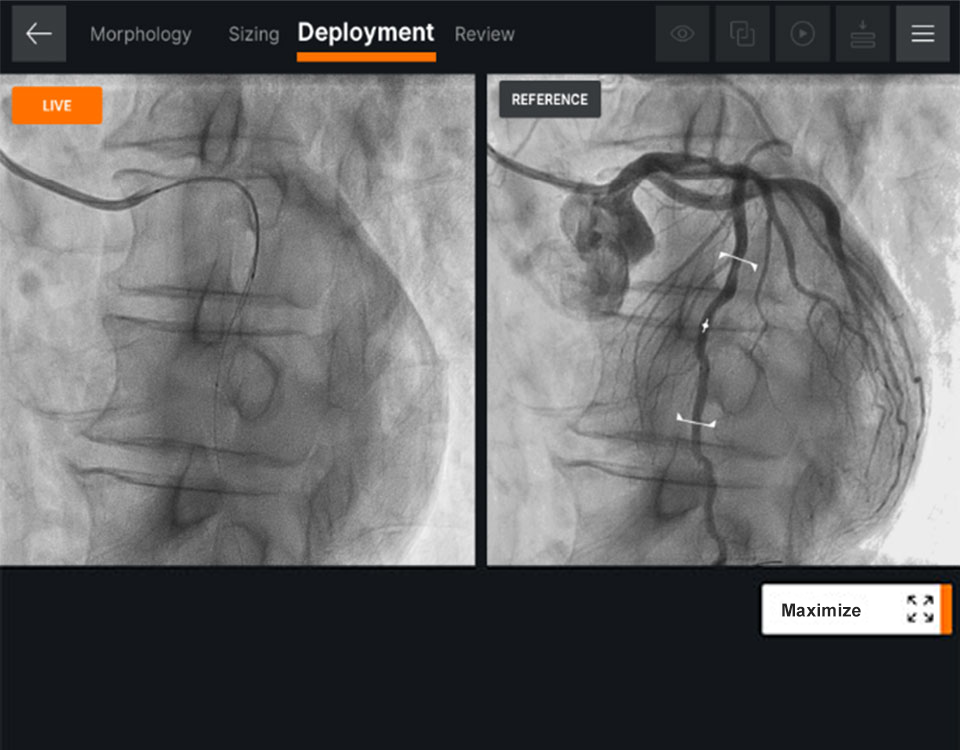
Ultreon™ 1.0 Software displays stent deployment screen with angio co‑registration
Ultreon™ 1.0 Software displays stent deployment screen with angio co‑registration
Post-PCI Guidance with Ultreon™ 1.0 Software
Review: Post-stent optimization2
Ultreon™ 1.0 Software displays instantaneous calculations of expansion and apposition values, reference lumen diameter and lumen diameter values in color-coded markers:
- Stent underexpansion indicator — orange
- Stent malapposition indicator — yellow
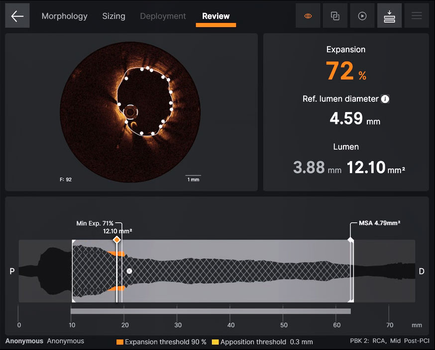
Ultreon™ 1.0 Software displays post-PCI review screen with stent expansion values
Ultreon™ 1.0 Software displays post-PCI review screen with stent expansion values
Malapposition Indicator2
Ultreon™ 1.0 Software helps ICs to quickly detect stent malapposition on the cross-sectional and lumen profile views:
- On the cross-sectional view, malapposed struts are indicated by yellow dots if the gap between the strut and the vessel wall exceeds the user’s preselected threshold setting.
- On the Lumen profile, malapposed zones are indicated by yellow bands when there’s a cluster of malapposed struts of length ≥ 1 mm and maximum circumferential angle ≥ 90 degrees
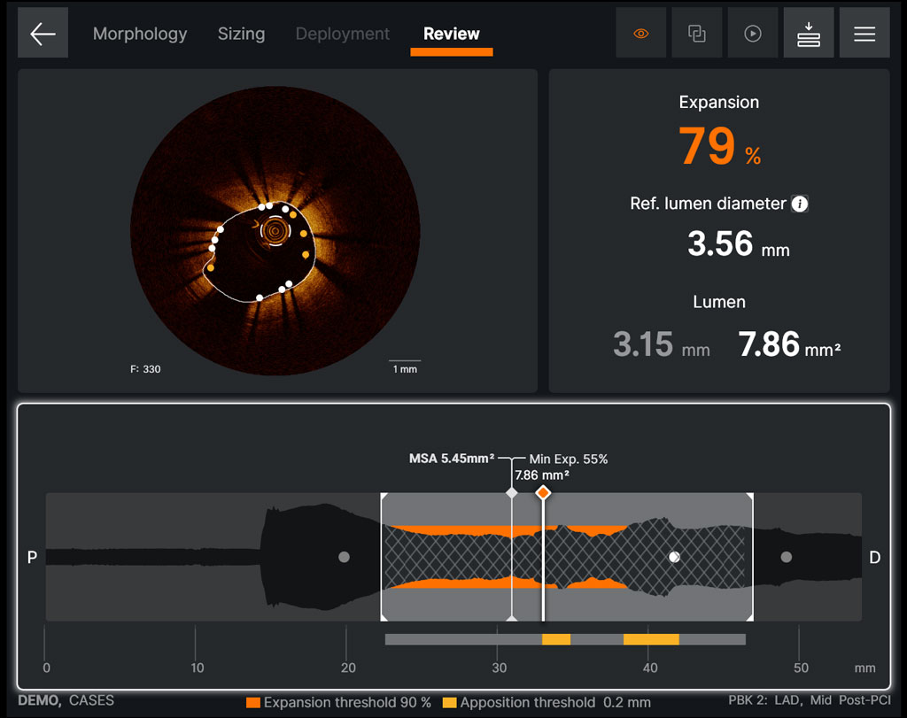
Ultreon™ 1.0 Software displays Malapposition Indicator on the Review screen
Ultreon™ 1.0 Software displays Malapposition Indicator on the Review screen
Pullback Comparison2
Physicians can quickly compare any two pullbacks by clicking on the Menu icon in the upper right corner. See the result of your PCI through vessel visualization pre- and post-stenting on one screen.
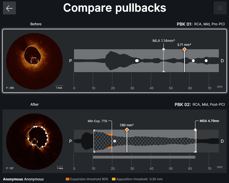
Ultreon™ 1.0 Software displays pullback comparison screen
Ultreon™ 1.0 Software displays pullback comparison screen
Watch How to Guide PCI with Ultreon™ 1.0 Software and MLD MAX Workflow:
Case-based presentation
References
- Data on File at Abbott.
- Ultreon™ 1.0 Software IFU. Refer to Instructions For Use (IFU) for additional information.
- Dean Kereiakes, Disrupt CAD III: Lithotripsy for Vessel Preparation in Calcified Coronary Arteries Prior to Stenting, CLD. Dec 29, 2020.
- Madhavan MV, Tarigopula M, Mintz GS, Maehara A, Stone GW, Généreux P. Coronary artery calcification: pathogenesis and prognostic implications. J Am Coll Cardiol. 2014 May 06;63(17):1703-14.
- Shlofmitz, E., Sosa, F.A., Ali, Z.A., Waksman, R., Jeremias, A., & Shlofmitz, R. (2019). OCT-Guided Treatment of Calcified Coronary Artery Disease: Breaking the Barrier to Stent Expansion. Current Cardiovascular Imaging Reports, 12(8), 32.
- Prati F, et al. Clinical impact of OCT findings during PCI: the CLI-OPCI II study. JACC: Cardiovasc Imaging. 2015;8(11):1297-1305. doi: 10.1016/j.jcmg.2015.08.013.
MAT-2104457 v3.0