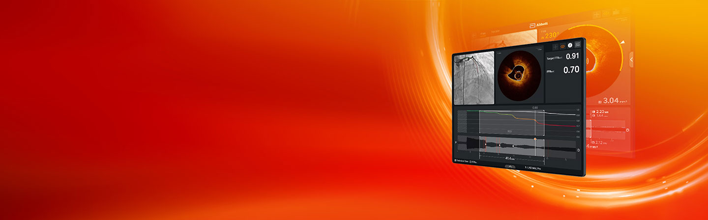
Imaging Reimagined with Ultreon™ 3.0 Software
High-resolution OCT imaging with physiology and AI-automated insights to enable simple, fast, and precise decisions for complex cases.


Delivers low-to-zero contrast OCT imaging at higher resolution than IVUS1
One-second Pullback
- Fast Pullback with streamlined setup and Al automated insights simplifies workflow
- Enables saline compatibility for renal risk patients
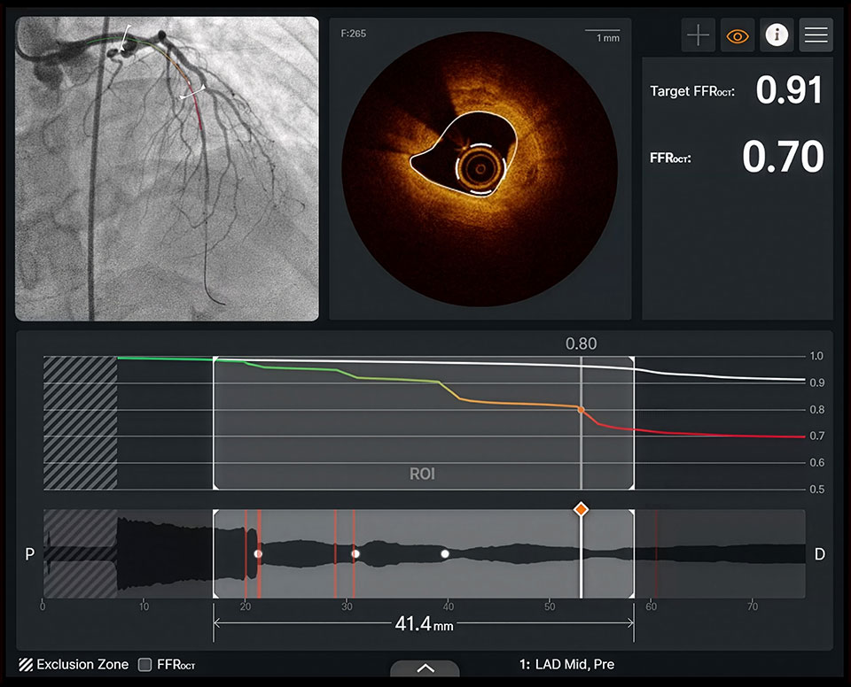
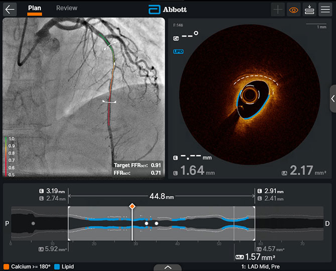
One Pullback, Total Perspective: Optimize flow recovery and predict PCI outcomes with FFROCT

FFROCT to optimize PCI
Evaluate the degree of ischemia for each lesion

Target FFROCT
Compare the physiologic impact of different treatment plans in real time

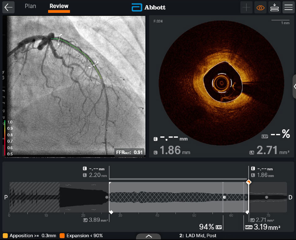
Post-PCI FFROCT
Predict patient outcomes, independent of MSA
Ultreon™ 3.0 Software with FFROCT is backed by data
82%
ACCURACY
Fusion Study
FFROCT demonstrated high accuracy compared to wire-based FFR2
0.70
HAZARD RATIO
FFROCT per 0.1 mm Hg/mm Hg
p=0.021
Illumien IV Substudy
Post-PCI FFROCT assessment is predictive of 2-year clinical outcomes independent of MSA3
Insights in seconds for faster procedure planning
Calcium
Arc and thickness automatically highlighted (orange) and measured
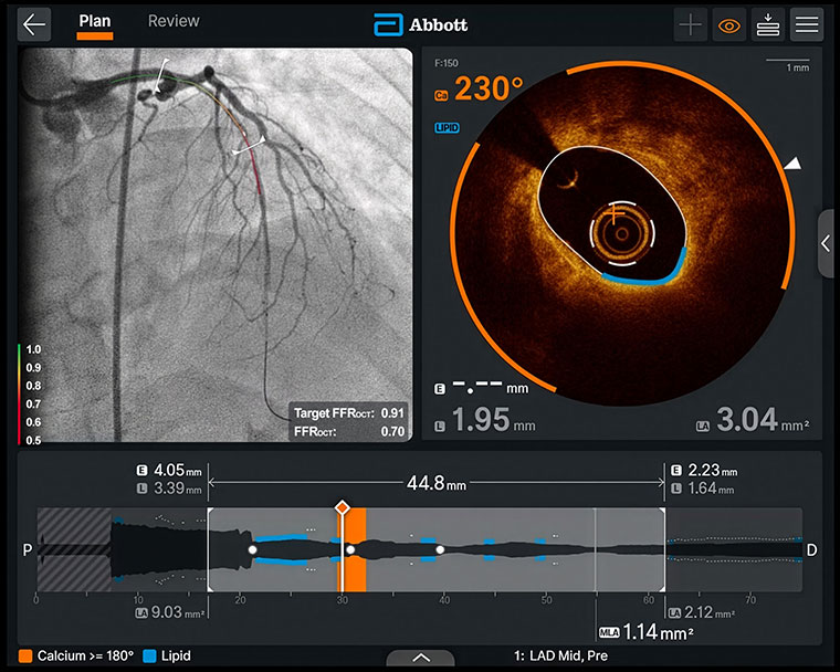
Lipid detection
Automatic detection (blue) helps you easily identify lipid-free landing zones
EEL
Automatically highlighted (dashed white line) and measured
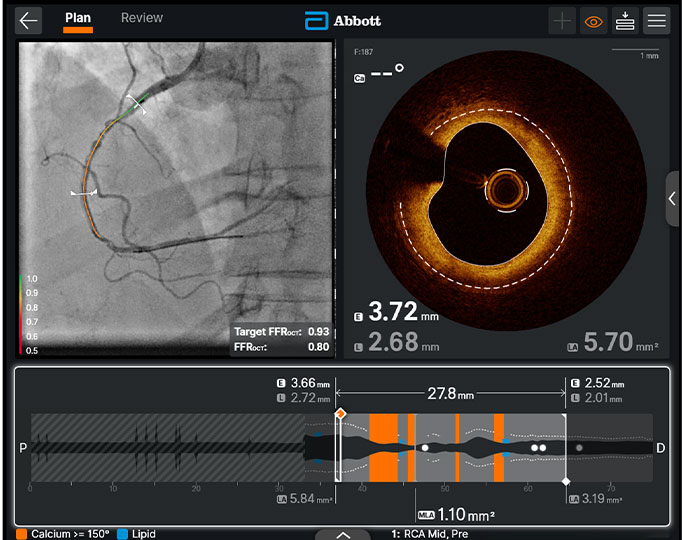
Automatic tri-registration
- Tri-registration of angiography, OCT, and physiology data
- Enables users to pinpoint the angiographic location of calcium, lipid, and flow-limiting lesions.

References
*FFROCT is the commercial name for Virtual Flow Reserve (VFR).
**Compared to previous Ultreon™ Software generations.
Ultreon™ 3.0 Software for OPTIS™ Next Imaging Systems Instructions for Use (IFU). Refer to IFU for additional information.
- Maehara A, Matsumura M, Ali ZA, et al. IVUS-Guided Versus OCT-Guided Coronary Stent Implantation: A Critical Appraisal. JACC: Cardiovascular Imaging. 2017;10(12):1487-1503.
- Jeremias A, et al. Optical Coherence Tomography–Based Functional Stenosis Assessment: FUSION—A Prospective Multicenter Trial. Circ Cardiovasc Interv. 2024:17:e013702.
- Johnson TW, et al. Impact of Optical Coherence Tomography-Based Post-PCI Physiology Assessment to Predict Clinical Outcomes: An ILUMIEN-IV Substudy. J Am Coll Cardiol. 2025 Jul 15;86(2):93-102.
MAT-2512693 v2.0