
Diagnostic catheters are used to navigate cardiac anatomy, capture critical cardiac information and deliver more precise therapy. Our diagnostic catheters are designed to help facilitate comprehensive data collection and meet your patients’ needs from simple to complex cases.
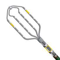
Advisor™ HD Grid X Mapping Catheter, Sensor Enabled™
The Advisor™ HD Grid X Mapping Catheter, Sensor Enabled™ allows you to place 16 electrodes where you need them, maneuvers bi-directionally within all chambers of the heart and offers a boost in accuracy, speed, and versatility1
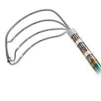
Advisor™ HD Grid Mapping Catheter, Sensor Enabled™
The Advisor™ HD Grid Mapping Catheter, Sensor Enabled™, is a first-of-its-kind, grid-patterned electrode configuration that offers high-density wave bipole recordings along and across the splines for direction independent mapping.2
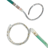
The Advisor™ VL and Advisor™ FL Mapping Catheter, Sensor Enabled™
The Advisor™ VL and FL Mapping Catheters, Sensor Enabled™ provides the power of choice. The Advisor™ VL Mapping Catheters, Sensor Enabled™ offers easy use in simple and straightforward mapping cases while the Advisor™ FL Mapping Catheters, Sensor Enabled™ precise model creation to facilitate complex arrhythmia diagnoses.3,4
References
- Abbott. Report on file. 91060435. A preclinical study with the objective of comparing Advisor HD Grid X Mapping Catheter, SE to Octaray and Advisor HD Grid Mapping Catheter, SE. Four participating independent physicians performed the study protocol with each of the HD Grid X Mapping Catheter, SE, Octaray, and Advisor HD Grid Mapping Catheter, SE catheters within the atria and ventricles of each of six swine study subjects. The protocol measured each catheter on (1) mapping and modeling speed; (2) model accuracy; and (3) ectopic burden.
- Abbott. Report on file. 90280703.
MAT-2007739 v3.0
Indications, Safety and Warnings
Advisor HD Grid X Mapping Catheter, Sensor Enabled
Rx Only. Brief Summary: Prior to using these devices, please review the Instructions for Use for a complete listing of indications, contraindications, warnings, precautions, potential adverse events, and directions for use.
United States: Required Safety Information
Indications for Use: The Advisor™ HD Grid X Mapping Catheter, Sensor Enabled™, is indicated for multiple electrode electrophysiological mapping of cardiac structures in the heart, i.e., recording or stimulation only. This catheter is intended to obtain electrograms in the atrial and ventricular regions of the heart. Contraindications: The catheter is contraindicated for patients with prosthetic valves, and patients with left atrial thrombus or myxoma, or interatrial baffle or patch via transseptal approach. This device should not be used with patients with active systemic infections. Patients unable to receive heparin or an acceptable alternative to achieve adequate anticoagulation. Warnings: Persons with a known history of allergies to any of the materials listed below may suffer an allergic reaction to this device. Before use, counsel the patient on the materials contained in the device and discuss a thorough history of allergies. This device contains: Acrylonitrilebutadienestyrene (ABS Cycolac) – Loctite Adhesive – Pellethane – Platinum Iridium alloy – Polyimide – Polyether block amide (PEBAX) – Polyethylene (High Density Polyethylene, HDPE) – Titanium. Cardiac catheterization procedures present the potential for significant xray exposure, which can result in acute radiation injury as well as increased risk for somatic and genetic effects, to both patients and laboratory staff due to the xray beam intensity and duration of the fluoroscopic imaging. Careful consideration must therefore be given for the use of this catheter in pregnant women. The safety and effectiveness of the device has not been established in pregnant women or prepubescent children. Careful consideration must therefore be given for the use of the device in pregnant women or prepubescent children. Catheter entrapment within the heart or blood vessels is a possible complication of electrophysiology procedures. Vascular perforation or dissection is an inherent risk of any electrode placement. Careful catheter manipulation must be performed in order to avoid device component damage, thromboembolism, cerebrovascular accident, cardiac damage, perforation, pericardial effusion, or tamponade. Risks associated with electrical stimulation may include, but are not limited to, the induction of arrhythmias, such as atrial fibrillation (AF), ventricular tachycardia (VT) requiring cardioversion, and ventricular fibrillation (VF). Do not use force to advance or withdraw catheter when resistance is encountered. Do not immerse the proximal handle or cable connector in fluids; electrical performance could be affected. Precautions: Maintain an activated clotting time (ACT) of greater than 300 seconds at all times during use of the catheter. This includes when the catheter is used in the right side of the heart. To prevent entanglement with concomitantly used catheters, use care when using the catheter in the proximity of the other catheters. Maintain constant irrigation to prevent coagulation on the distal paddle. Inspect irrigation tubing for obstructions, such as kinks and air bubbles. If irrigation is interrupted, remove the catheter from the patient and inspect the catheter. Ensure that the irrigation ports are patent and flush the catheter prior to reinsertion. Use the straightener during the insertion process to avoid damage to the hemostasis valve. Always straighten the catheter before insertion or withdrawal. Catheter advancement must be performed under fluoroscopic guidance to minimize the risk of cardiac damage, perforation, or tamponade. Compatible navigation and real time visualization systems may also be considered. Do not use if the catheter appears damaged, kinked, or if there is difficulty in deflecting the distal section to achieve the desired curve. Catheter materials are not compatible with magnetic resonance imaging (MRI). One or more components of this device may contain the following substance defined as CMR 1B in a concentration above 0.1% weight by weight: Nmethyl2pyrrolidone (NMP): Chemical Abstracts Service (CAS) No. 872504; EC No. 2128281. Based on a quantitative toxicological assessment it has been determined that NMP released from this device is unlikely to cause adverse reproductive effects. Potential Adverse Events: Complications related to the use of the device include, but are not limited to, the following: New or worsening of existing arrhythmia including Atrial fibrillation, Ventricular tachycardia requiring cardioversion, Supraventricular tachycardia (SVT) and Ventricular fibrillation Cardiac perforation including Pericardial effusion or Cardiac tamponade Bleeding including access site Hemorrhage / bleeding, Ecchymosis and Hematoma Vascular access complications or peripheral vascular injury including Femoral artery dissection, Dissection, Arteriovenous fistula and Pseudoaneurysm formation Pulmonary vein stenosis, Heart failure, Volume overload, Hypotension, Embolism, Cerebrovascular accident (CVA)/stroke, Infection, Pneumonia, Pulmonary edema, Immunological reaction, Pain and Pericarditis.
MAT-2503929 v1.0
Advisor HD Grid Mapping Catheter, Sensor Enabled
Indications: The Advisor™ HD Grid Mapping Catheter, Sensor Enabled™, is indicated for multiple electrode electrophysiological mapping of cardiac structures in the heart, i.e., recording or stimulation only. This catheter is intended to obtain electrograms in the atrial and ventricular regions of the heart.
Contraindications: Contraindicated for patients with prosthetic valves and patients with left atrial thrombus or myxoma, or interatrial baffle or patch via transseptal approach. This device should not be used with patients with active systemic infections. The catheter is contraindicated in patients who cannot be anticoagulated or infused with heparinized saline.
Caution: This product is intended for use by or under the direction of a physician. Prior to use, reference Instructions for Use, inside the product carton (when available) or at eifu.abbottvascular.com or at manuals.sjm.com for more detailed information on Indications, Contraindications, Warnings, Precautions and Adverse Events.
Warnings: Cardiac catheterization procedures present the potential for significant x-ray exposure, which can result in acute radiation injury as well as increased risk for somatic and genetic effects, to both patients and laboratory staff due to the x-ray beam intensity and duration of the fluoroscopic imaging. Careful consideration must therefore be given for the use of this catheter in pregnant women. Catheter entrapment within the heart or blood vessels is a possible complication of electrophysiology procedures. Vascular perforation or dissection is an inherent risk of any electrode placement. Careful catheter manipulation must be performed in order to avoid device component damage, thromboembolism, cerebrovascular accident, cardiac damage, perforation, pericardial effusion, or tamponade. Risks associated with electrical stimulation may include, but are not limited to, the induction of arrhythmias, such as atrial fibrillation (AF), ventricular tachycardia (VT) requiring cardioversion, and ventricular fibrillation (VF). Catheter materials are not compatible with magnetic resonance imaging (MRI).
Precautions: Maintain an activated clotting time (ACT) of greater than 300 seconds at all times during use of the catheter. This includes when the catheter is used in the right side of the heart. To prevent entanglement with concomitantly used catheters, use care when using the catheter in the proximity of the other catheters. Maintain constant irrigation to prevent coagulation on the distal paddle. Inspect irrigation tubing for obstructions, such as kinks and air bubbles. If irrigation is interrupted, remove the catheter from the patient and inspect the catheter. Ensure that the irrigation ports are patent and flush the catheter prior to re-insertion. Always straighten the catheter before insertion or withdrawal. Do not use if the catheter appears damaged, kinked, or if there is difficulty in deflecting the distal section to achieve the desired curve. Do not use if the catheter does not hold its curve and/or if any of the irrigation ports are blocked. Catheter advancement must be performed under fluoroscopic guidance to minimize the risk of cardiac damage, perforation, or tamponade.
MAT-2007568 v2.0
Advisor VL Circular Mapping Catheter, Sensor Enabled
Rx Only. Brief Summary: Prior to using these devices, please review the Instructions for Use for a complete listing of indications, contraindications, warnings, precautions, potential adverse events, and directions for use.
United States: Required Safety Information
Indications: The Advisor™ VL Circular Mapping Catheter, Sensor Enabled™ is a steerable electrophysiology catheter with integrated sensors. The catheter is used for recording intracardiac signals and cardiac stimulation during diagnostic electrophysiology studies. The catheter can be used to map the atrial regions of the heart. Contraindications: The catheter is contraindicated for patients with prosthetic valves and patients with left atrial thrombus or myxoma, or interatrial baffle or patch via transseptal approach. This device should not be used via retrograde approach. This device is not recommended for use in the ventricles. The device is not intended for transcatheter ablation. This device should not be used with patients with active systemic infections. Warnings: Cardiac catheterization procedures present the potential for significant x-ray exposure, which can result in acute radiation injury as well as increased risk for somatic and genetic effects, to both patients and laboratory staff due to the x-ray beam intensity and duration of the fluoroscopic imaging. Careful consideration must therefore be given for the use of this catheter in pregnant women. Catheter entrapment within the heart or blood vessels is a possible complication of electrophysiology procedures. To unentangle the catheter, fully open the loop and straighten the catheter shaft, then rotate the handle clockwise. Vascular perforation or dissection is an inherent risk of any electrode placement. Careful catheter manipulation must be performed in order to avoid thromboembolism, cerebral accident, cardiac damage, perforation, pericardial effusion, or tamponade. Risks associated with electrical stimulation may include, but are not limited to, the induction of arrhythmias, such as atrial fibrillation (AF), ventricular tachycardia (VT) requiring cardioversion, and ventricular fibrillation (VF). Precautions: Maintain an activated clotting time (ACT) of greater than 300 seconds at all times during use of the catheter. Excessive bending or kinking of the catheter may cause damage to the catheter. Always straighten the catheter and open the loop before insertion or withdrawal. Catheter advancement must be performed under fluoroscopic guidance to minimize the risk of cardiac damage, perforation, or tamponade. Compatible navigation and visualization systems may be used in conjunction with fluoroscopy.
Advisor FL Circular Mapping Catheter, Sensor Enabled
Rx Only. Brief Summary: Prior to using these devices, please review the Instructions for Use for a complete listing of indications, contraindications, warnings, precautions, potential adverse events, and directions for use.
United States: Required Safety Information
Indication: The Advisor™ FL Circular Mapping Catheter, Sensor Enabled™ is steerable electrophysiology catheter with integrated sensors. The catheter is used for recording intracardiac signals and cardiac stimulation during diagnostic electrophysiology studies. The catheter can be used to map the atrial regions of the heart. Contraindications: The catheter is contraindicated for patients with prosthetic valves and patients with left atrial thrombus or myxoma, or interatrial baffle or patch via transseptal approach. This device should not be used via retrograde approach. This device is not recommended for use in the ventricles. The device is not intended for transcatheter ablation.This device should not be used with patients with active systemic infections. Warnings: Cardiac catheterization procedures present the potential for significant x-ray exposure, which can result in acute radiation injury as well as increased risk for somatic and genetic effects, to both patients and laboratory staff due to the x-ray beam intensity and duration of the fluoroscopic imaging. Careful consideration must therefore be given for the use of this catheter in pregnant women. Catheter entrapment within the heart or blood vessels is a possible complication of electrophysiology procedures. Vascular perforation or dissection is an inherent risk of any electrode placement. Careful catheter manipulation must be performed in order to avoid thromboembolism, cardiac damage, perforation, or tamponade. The induction of atrial fibrillation (AF), ventricular tachycardia (VT) requiring cardioversion, and ventricular fibrillation (VF) can be risks associated with electrical stimulation. Precautions: Excessive bending or kinking of the catheter may cause damage to the catheter. Always straighten the catheter before insertion or withdrawal. Catheter advancement must be performed under fluoroscopic guidance to minimize the risk of cardiac damage, perforation, or tamponade. Compatible navigation and visualization systems may be used in conjunction with fluoroscopy.
MAT-2501377 v1.0