
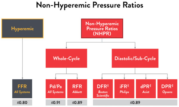
RFR – Resting Full-cycle Ratio(安静時の全心周期における圧較差指標)
アボットのPressureWire™ X ガイドワイヤーは、臨床状況に応じてFFRの代替として測定できる安静時指標(RFR)の測定も可能です1。FFRと同様に、RFRは心外膜血管における狭窄の機能的重症度を評価することができます。FFRとは異なり、RFRはアデノシンなどの血管拡張薬による負荷を必要としない安静時における指標です1。
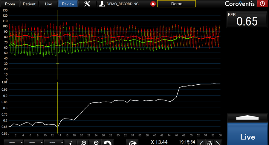
RFRの測定
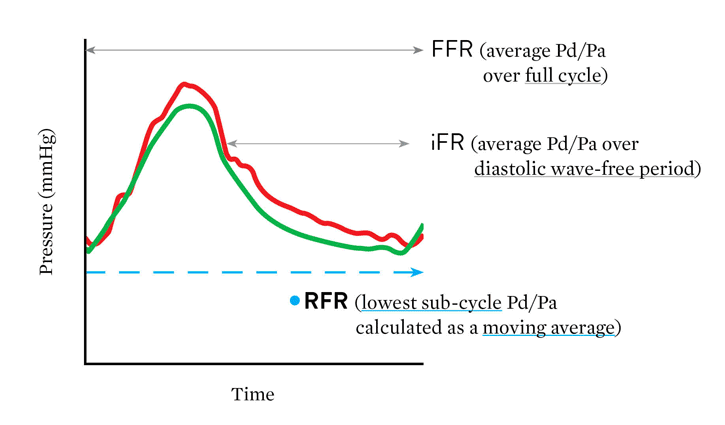
iFR‡(瞬時血流予備量比)と臨床的に同等であるアボットのRFR(安静時の全心周期における圧較差指標)は、全心周期における最小のPd/Paを1心拍ごとに求め、それらを5心拍平均で算出します。iFR‡ や他の安静時指標 とは異なり、RFR は心周期全体における Pd/Pa の最小値から計算されます。
RFRの利点
「iFR‡ に対する RFR の主な利点は、拡張期に限定した特定の時相を選択していないことです。RFRは心周期全体の最小 Pd/Pa を計算することにより、安静時における狭窄前後の最大圧勾配を導き出しています。」
– Lee, et al. Circulation 2019
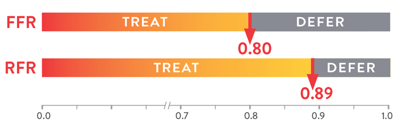
RFRのカットオフ値は0.89です1,3,4。
- FFRが≤0.89の場合、経皮的冠動脈インターベンション(PCI)が遠隔期のMIと緊急血行再建の発生を低減させたことが報告されています
- FFRが>0.89場合、一般に血行再建の対象にはなりません
他の安静時指標とRFRを比較して
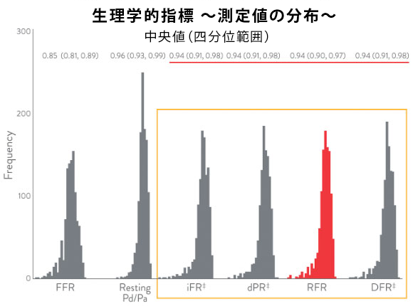
すべての安静時指標は互いに密接に相関しており、血行再建の可否に対して同等の識別能を有することが証明されています2。 IRIS-FFRレトロスペクティブ研究(1,506例、1,833病変)では、5 つの安静時指標(安静時 Pd/Pa、iFR‡、RFR、dPR‡、および DFR‡)に基づいて血行再建が延期された症例の予後が調査され、すべての安静時指標間で同等の結果をもたらすと結論付けられました4。
それぞれの安静時指標のカットオフ値
RFRの主な研究結果
RFRは、2,500例以上、3,500病変以上を対象に研究されています。
| 主なRFR研究 | ||
|---|---|---|
| 病変数 | 患者数 | |
| RE-VALIDATE RFR6 | 501 | 431 |
| VALIDATE RFR1 | 672 | 504 |
| ILUMIEN I + PREDICT3 | 313 | 299 |
| IRIS-FFR7 | 1,102 | 926 |
| 3V FFR-FRIENDS2 | 1,024 | 435 |
| 計 | 3,612 | 2,595 |
FFRと安静時指標(RFR)の使用が有効なケース8
| 安静時指標(RFR)の使用が有効なケース | ||
|---|---|---|
| 安静時指標 | FFR | |
| タンデム病変9 | +++ | + |
| びまん性病変10 | +++ | + |
| 左前下行枝11 | +++ | ++ |
| 急性冠症候群12 | +++ | ++ |
| 同等に有効なケース | ||
| 多枝病変13 | +++ | +++ |
| 糖尿病14 | +++ | +++ |
| FFRの使用が有効なケース | ||
| 左主幹部病変15 | – | +++ |
| 左室機能障害16 | – | +++ |
| PCI 後の側枝評価17 | – | +++ |
| PCI後の評価18 | – | +++ |
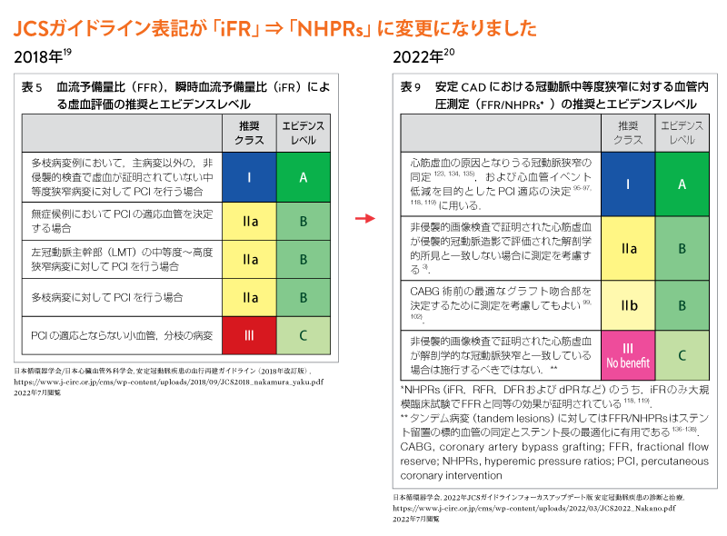
日本循環器学会におけるガイドライン

‡ 第三者企業またはそれぞれの所有者の商標を示します。
References
- Svanerud J, Ahn JM, Jeremias A, et al. Validation of a novel non-hyperaemic index of coronary artery stenosis severity: the Resting Full-cycle Ratio (VALIDATE RFR) study. EuroIntervention. 2018;14:806-814.
- Lee JM, Choi KH, Park J, et al. Physiological and clinical assessment of resting physiological indexes. Circulation. 2019;139:889-900. doi: 10.1161/CIRCULATIONAHA.118.037021.
- Jeremias A, et al. Resting full-cycle ratio (RFR): a novel physiologic index compared to Fractional Flow Reserve (FFR) in assessing the hemodynamic severity of a coronary stenosis: ILUMIEN I + PREDICT. EuroPCR 2018.
- Ahn JM, et al. IRIS FFR: prognostic performance of five resting pressure-derived indexes of coronary physiology. TCT 2018.
- Ali ZA. Invasive assessment of coronary physiology is here to stay. TCT 2019.
- Kumar G, Desai R, Goreet A, al. RE-VALIDATE: REal world VALIDATion of the non-hyperemic InDex of coronary Artery sTEnosis severity: resting full-cycle ratio (RFR) – RE-VALIDATE RFR. CRT 2019.
- Data on file at Abbott.
- Ali Z. Invasive Assessment of Coronary Physiology Is Here to Stay! TCT 2019.
- Kikuta Y, Cook CM, Sharp ASP, et al. Pre-angioplasty instantaneous wave-free ratio pullback predicts hemodynamic outcome In humans with coronary artery disease: primary results of the International MulticenteriFR GRADIENT Registry. J Am Coll Cardiol Intv. 2018;11:757-767.
- Jeremias A, Davies JE, Maehara A, et al. Blinded physiological assessment of residual ischemia after uccessful angiographic percutaneous coronary intervention: the DEFINE PCI study. JACC Cardiovasc Interv. 2019;12(20):1991-2001.
- Sen S, Ahmad Y, Dehbi HM, et al. Clinical events after deferral of LAD revascularization following physiological coronary assessment. J Am Coll Cardiol. 2019;73(4):444-453.
- Escaned J, Ryan N, Mejía-Rentería H, et al. Safety of the deferral of coronary revascularization on the basis of instantaneous wave-free ratio and fractional flow reserve measurements in stable coronary artery disease and acute coronary syndromes. JACC Cardiovasc Interv. 2018;11(15):1437-1449.
- Escaned J, Collet C, Ryan N, et al. Clinical outcomes of state-of-the-art percutaneous coronary revascularization in patients with de novo three vessel disease: 1-year results of the SYNTAX II study. Eur Heart J. 2017;38(42):3124-3134.
- DEFINE-FLAIR Trial Investigators. Comparison of major adverse cardiac events between instantaneous wave-free ratio and fractional flow reserve–guided strategy in patients with or without type 2 diabetes. JAMA Cardiol. 2019;4(9):857-864.
- Mallidi J, Atreya AR, Cook J, et al. Long-term outcomes following fractional flow reserve-guided treatment of angiographically ambiguous left main coronary artery disease: a meta-analysis of prospective cohort studies. Catheter Cardiovasc Interv. 2015;86(1):12-18.
- Di Gioia G, De Bruyne B, Pellicano M, et al. Fractional flow reserve in patients with reduced ejection fraction. Eur Heart J. 2020;41(17):1665-1672.
- Koo BK, Kang HJ, Youn TJ, et al. Physiologic assessment of jailed side branch lesions using fractional flow reserve. J Am Coll Cardiol. 2005;46(4):633-637.
- Johnson NP, Tóth GG, Lai D, et al. Prognostic value of fractional flow reserve: linking physiologic severity to clinical outcomes. J Am Coll Cardiol. 2014;64(16):1641-1654. doi: 10.1016/j.jacc.2014.07.973.
- 日本循環器学会/日本心臓血管外科学会. 安定冠動脈疾患の血行再建ガイドライン(2018年改訂版). https://www.j-circ.or.jp/cms/wp-content/uploads/2018/09/JCS2018_nakamura_yaku.pdf 2022年7月閲覧
- 日本循環器学会. 2022年JCSガイドラインフォーカスアップデート版 安定冠動脈疾患の診断と治療. https://www.j-circ.or.jp/cms/wp-content/uploads/2022/03/JCS2022_Nakano.pdf 2022年7月閲覧
MAT-2304625 v1.0