OPTIS™ Next Imaging Systems
OCT imaging and coronary physiology on one platform
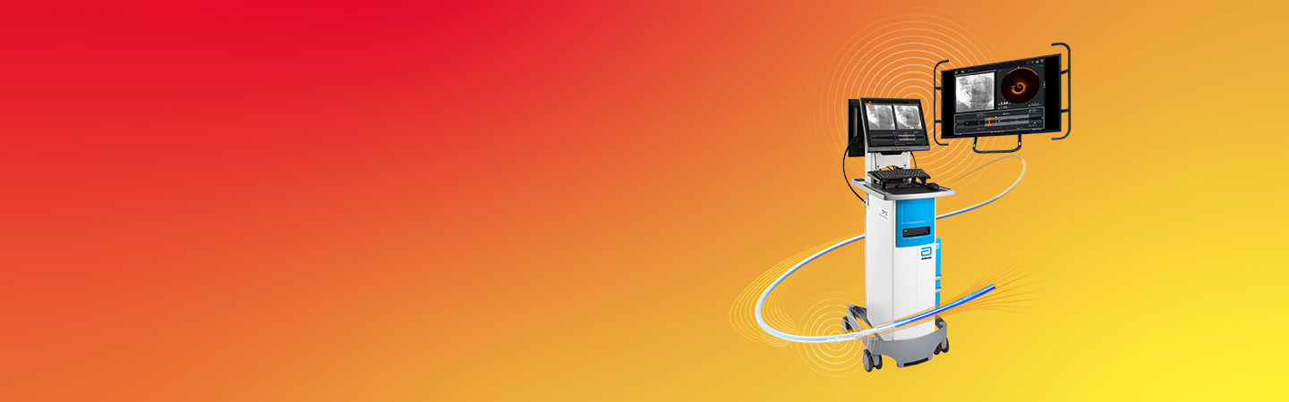
OPTIS™ Next Imaging Systems offer high resolution OCT intravascular imaging and coronary physiology (RFR/FFR) on one platform to guide PCI with informed decisions. The OPTIS™ Next Imaging Systems - Integrated System and Mobile System - powered by Ultreon™ Software which includes coronary physiology measurement, used with the Dragonfly OpStar™ Imaging Catheter, represent our intravascular imaging products that are designed for imaging of coronary arteries.
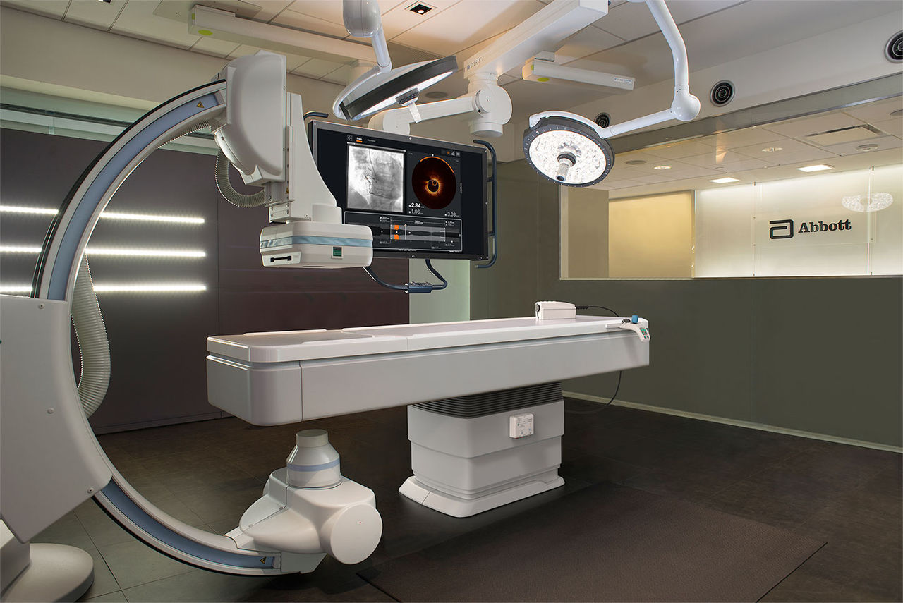
OPTIS™ Integrated Next System set up. Components shown include Wi-Box™ AO Transmitter, DOC, TSC and OCT user interface displayed on Boom Monitor
OPTIS™ Integrated Next System set up. Components shown include Wi-Box™ AO Transmitter, DOC, TSC and OCT user interface displayed on Boom Monitor
When interventional cardiologists (ICs) have access to sophisticated OCT imaging and coronary physiology in the cath lab, data show improved outcomes during PCI stent procedures:1-3
The OPTIS™ Next Imaging Systems are seamlessly integrated into the cath lab and PCI workflow. Benefits include:
Access to OCT and RFR/FFR on one system for informed PCI decision-making
- The OPTIS™ Next Imaging Systems enable OCT imaging and coronary physiology measurement using resting full-cycle ratio (RFR) and fractional flow reserve (FFR). This integration provides a versatile system for the multiple modalities used during PCI, reducing set-up time and eliminating clutter from multiple cables and components.
Automated OCT software features inform aspects of PCI planning:
- Rapid image processing: The OPTIS™ Next Imaging Systems have a less than three second pullback speed enabling fast image acquisition and immediate interpretation of lesion morphology. Images appear on the OCT interface screen right after the pullback.
- Simultaneous angio and OCT co-registration: OCT software provides real time instantaneous synchronization of angiographic and OCT images for side-by-side viewing to help physicians clearly:
1) Identify stenotic lesion(s)
2) Mark stenotic locations to facilitate accurate stent placement - Post-PCI optimization information on one screen
The OCT software provides post-PCI information on one screen (apposition and expansion indicators, dissection visualization) to guide a user step-by-step to optimize PCI to ensure adequate stent expansion.
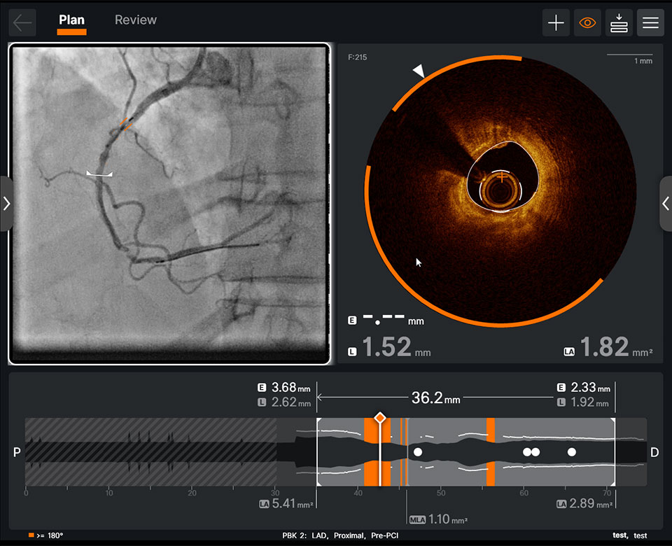
Pre-PCI OCT: Integration of AI-powered insights to your angiography panel helps you see calcium simply
Pre-PCI OCT: Integration of AI-powered insights to your angiography panel helps you see calcium simply
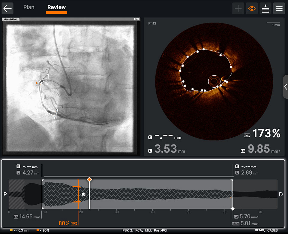
Post-PCI OCT: The OCT software provides post-PCI information on one screen
Post-PCI OCT: The OCT software provides post-PCI information on one screen
Easy integration in the cath lab for workflow efficiency with fewer cables and less clutter
-
Cath Lab Systems Integration
OPTIS™ Next Imaging Systems are designed to meet your specific cath lab needs for OCT-guided PCI and are easy to set up in your cath lab. Learn more about cath lab systems integration.
-
Ultreon™ Software
Ultreon™ Software is an imaging platform that uses the power of artificial intelligence (AI) to analyze lesion data and present unique OCT and angiographic insights so physicians can make fast and accurate decisions to achieve optimal patient results.

References
OPTIS™ Integrated Next Imaging System Instructions for Use (IFU). OPTIS™ Mobile Next IFU. Ultreon™ 2.0 Software IFU. Refer to IFUs for additional information.
- Jones DA, et al Angiography alone versus angiography plus optical coherence tomography to guide percutaneous coronary intervention. JACC: Cardiovasc Interv. 2018;11(14):1313-1321. doi: 10.1016/j.jcin.2018.01.274.
- Pijls, N., et al. 2-year follow-up of the FAME (Fractional Flow Reserve Versus Angiography for Multivessel Evaluation) study. JACC Vol. 56, No. 3, 2010.
- Ahn JM, et al. IRIS FFR: prognostic performance of five resting pressure-derived indexes of coronary physiology. TCT 2018.
- Z Ali et al., Optical Coherence Tomography–Guided versus Angiography-Guided PCI, NEJM, DOI: 10.1056/NEJMoa230586.
- N.R. Holm et al., OCT or Angiography Guidance for PCI in Complex Bifurcation Lesions, NEJM, DOI: 10.1056/NEJMoa2307770 (OCTOBER).
- Bergmark, B. et al: Decision-Making During Percutaneous Coronary Intervention Guided by Optical Coherence Tomography: Insights From the LightLab Initiative. Circ Cardiovasc Interv. 2022;15:e011851. DOI: 10.1161/CIRCINTERVENTIONS.122.011851.
MAT-2103698 v2.0