Cath Lab System Integration with OPTIS™ Next Imaging Systems
OPTIS™ Next Imaging Systems are easily integrated with the cath lab systems.
Integration of FFR/RFR with cath lab recording hemodynamic system via Wi‑Box™ AO Transmitter connection
The OPTIS™ Next Imaging System displays coronary physiology (FFR/RFR) measurement using a wireless PressureWire™ X Guidewire and an external aortic pressure (AO) transducer. Aortic pressure signal is sent from an AO transducer to Wi-Box™ AO Transmitter and wirelessly transmitted to the OPTIS™ Next Imaging System.1
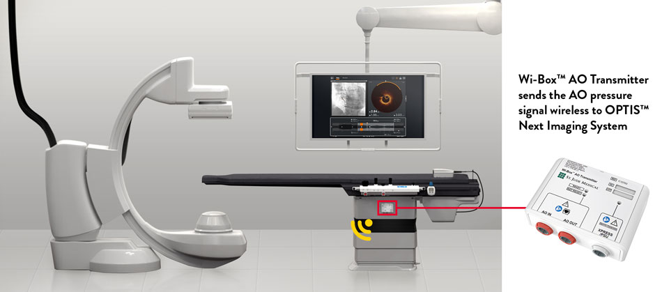
Wi-Box™ AO Transmitter sends signal wireless to the OPTIS™ Integrated Next System. Components shown include Wi-Box™ AO Transmitter, DOC, tableside controller (TSC) and OCT user interface display on the Boom Monitor
Wi-Box™ AO Transmitter is a small device easily installed by mounting on the base of the cath lab table. No cables to connect to OPTIS™ Next Imaging System, no clutter, no interference with the flow of the PCI procedure. Wi-Box™ AO Transmitter sends the AO pressure signal wirelessly to the OPTIS™ Next Imaging System and the output is displayed on the existing cath lab monitors.
DICOM‡ Integration
OPTIS™ Next Imaging Systems implement several DICOM‡ services for exchanging information and images. These systems allow for the querying of patient information from a network storage device, as well as the storing of acquired OCT images and physiology images, as “secondary capture” images, to the network storage device or CD/DVD.
DICOM‡ integration features:2
- DICOM‡ Modality Worklist Query (results shown in Add Patient wizard)
- Export to DICOM‡ storage server
- Export to USB (DICOM‡)
- Export to USB (native)
To view DICOM‡ conformance statement, please click here.
DICOM‡=Digital Imaging and Communication in Medicine
Connection to the Boom monitor
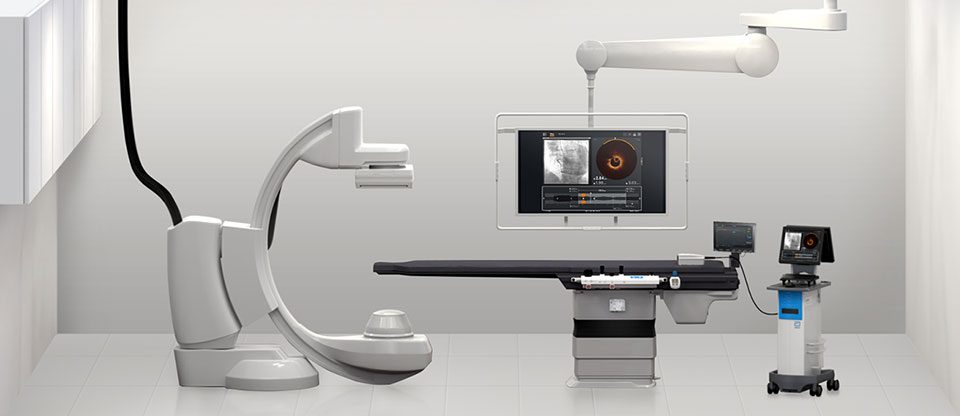
OPTIS™ Next Imaging Systems can be easily connected to standard Boom monitors in the cath lab to display the OCT user interface on the Boom monitor to view PCI information from the procedure table.
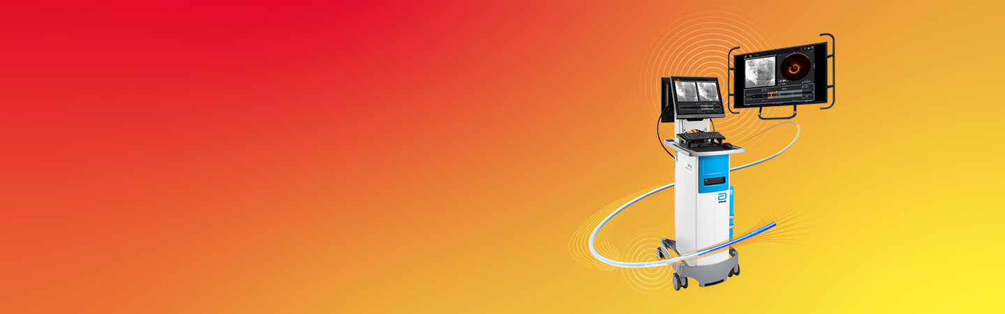
OPTIS™ Mobile Next System and an added Ultiri™ Measurement System set up. Components shown include Wi-Box™ AO Transmitter, DOC, TSC and OCT user interface displayed on the Boom Monitor.
Connection options:
- OPTIS™ Integrated Next Imaging System is connected to Boom Monitor via DVI Extender
- OPTIS™ Mobile Next System is connected via Connectivity Box
Connection to cath lab angio systems
The OPTIS™ Next Imaging Systems can be integrated with the cath lab angio systems to display OCT and angio co-registration (ACR) on the same screen.
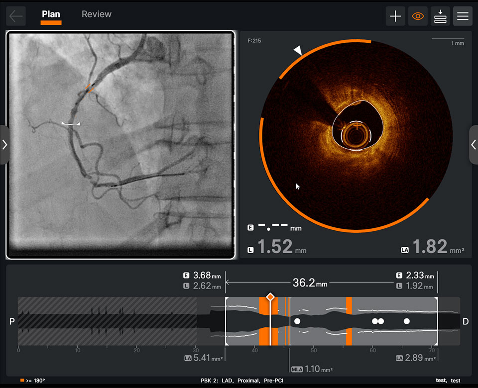
Integration of AI-powered insights to your angiography panel helps you see calcium simply
Integration of AI-powered insights to your angiography panel helps you see calcium simply
Notes
- Compatible with most hemodynamic systems in the cath lab that are following ANSI/AAMI standards.
- Available with Ultreon™ Imaging Software.
OPTIS™ Integrated Next Imaging System Instructions for Use (IFU). OPTIS™ Mobile Next IFU. Ultreon™ 2.0 Software IFU. Refer to IFUs for additional information.
MAT-2104275 v2.0