
Epicardial Assessment with FFR, RFR, and PPG
Epicardial physiology indices such as Fractional Flow Reserve (FFR), Resting Full-cycle Ratio (RFR) and Pullback Pressure Gradient (PPG) are utilized to determine the need and predict the likelihood of successful percutaneous coronary intervention (PCI).
FFR or RFR assessments are performed to assess the functional significance of epicardial coronary lesions at stress or rest, respectively.1 PPG is a novel epicardial index that objectively quantifies focal epicardial disease to identify patients that are most likely to benefit from PCI.
Measure FFR, RFR, and PPG with PressureWire™ X Guidewire, the world's only2,3 wireless physiology wire with a hydrophilic-coated3 design for exceptional handling performance and accurate, reliable readings.4,5

FFR: Fractional Flow Reserve
Measuring FFR
FFR is a measure of blood pressure difference across a coronary artery stenosis to determine if a stenosis is physiologically significant, which can impede oxygen delivery to the heart muscle.1
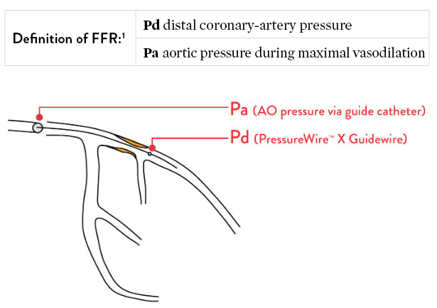
FFR is the gold standard of assessing a functionally significant coronary stenosis.6,7
Why Use FFR?
34%
of the time angiography is inaccurate
Coronary angiography is not accurate in guiding the functional significance of stenosis in 34% of cases, compared to FFR.8
35%
risk reduction in death/MI with FFR
FFR-guided percutaneous coronary intervention (PCI) leads to a 35% risk reduction in death and myocardial infarction (MI), compared to relying only on angiography.9
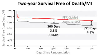
62%
relative risk reduction in MACCE
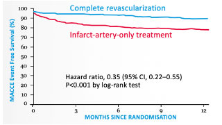
Complete revascularization with PressureWire™ X Guidewire led to a 62% relative risk reduction in major adverse cardiac and cerebrovascular events (MACCE) at 1 year.10 The DANAMI-3-PRIMULTI trial also revealed that untreated “FFR-positive” non-infarct-related artery lesions are associated with a 31% incidence of MACCE at 1 year.10
RFR: Resting Full-Cycle Ratio
PressureWire™ X Guidewire measures RFR, a resting index that can be measured as an alternative to FFR depending on the clinical situation.11 Like FFR, RFR can identify functionally significant epicardial coronary stenosis. Unlike FFR, RFR is a non-hyperemic index that does not require the administration of a vasodilator such as adenosine.11
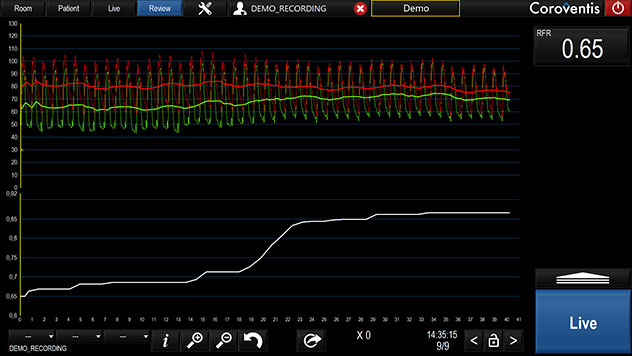
Measuring RFR
Clinically equivalent to the instantaneous wave-free ratio (iFR‡) resting index, Abbott's RFR resting index scans through diastole and systole for the lowest pd/pa ratio. Unlike iFR‡ or diastolic pressure ratio (dPR‡), RFR is calculated from the lowest value of Pd/Pa over the entire cardiac cycle.12
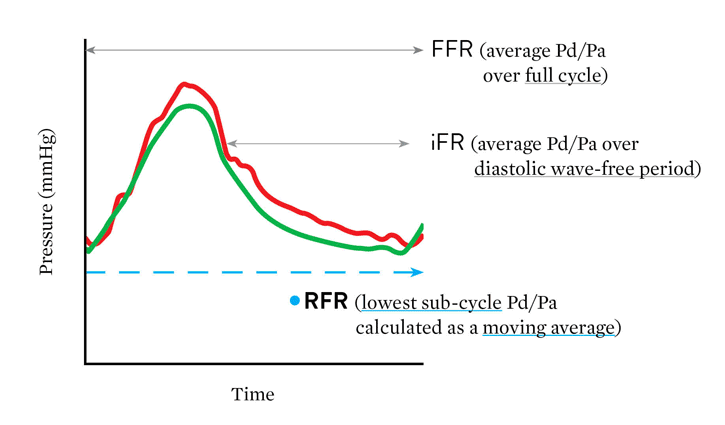
Advantage of RFR
"The major advantage of RFR over iFR‡ is that RFR does not require identification of a specific landmark or selection of a specific time point during diastole. By calculating the minimum Pd/Pa over the entire cardiac cycle, RFR calculates the maximum pressure gradient across the stenosis during resting status."
– Lee, et al. Circulation 2019
The cutoff value of RFR is 0.89:11,13,14
- When RFR ≤ 0.89, treatment with percutaneous coronary intervention (PCI) may be beneficial
- When RFR > 0.89, deferral of PCI may be beneficial
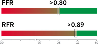
Green indicates deferral of PCI may be beneficial, and red indicates a treatment of PCI may be beneficial.
Assessing Serial or Diffuse Lesions with RFR Pullback
In patients with multiple sequential stenoses or diffuse lesions, pullback pressure assessment can identify lesions with functional significance. RFR can be calculated along the entire length of a stenotic vessel using pullback, which measures pressure gradient at any given location.11
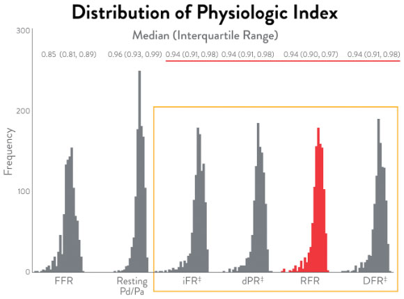
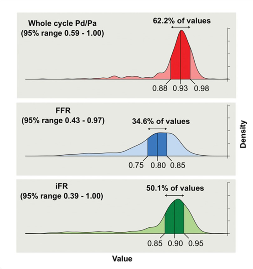
RFR Compared to Other Resting Indices
All resting pressure-derived indices closely correlate with one another, revealing the same discriminatory ability to guide intervention.12 The large IRIS-FFR retrospective study (1,506 patients, 1,833 lesions) examined deferred lesion failure among 5 resting indices—resting Pd/Pa, iFR‡, RFR, dPR‡, and DFR‡—and concluded that all resting indices had similar outcomes in deferred lesions.14
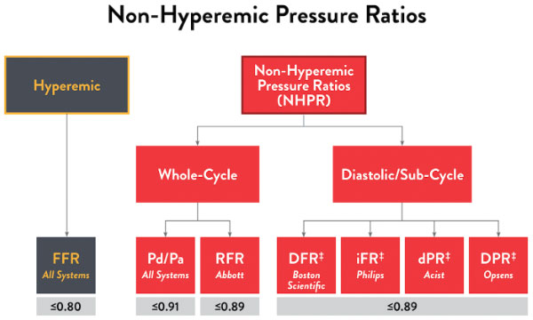
Here are the cutoff values for the various non-hyperemic pressure ratios.16
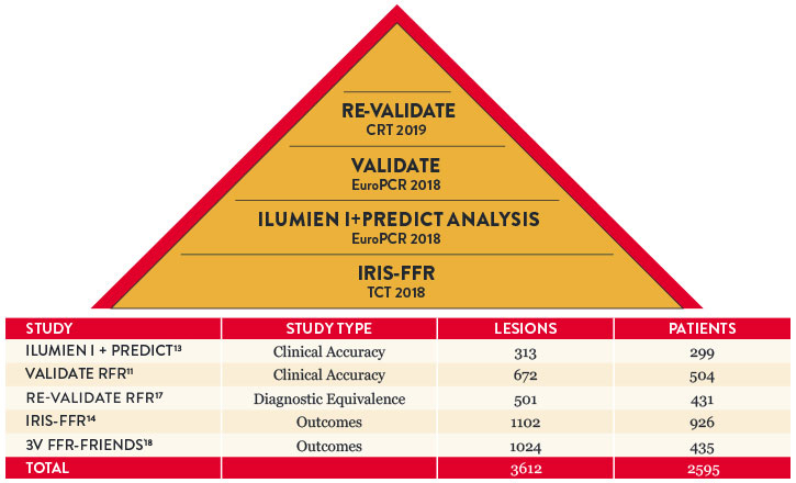
Key RFR Findings
RFR has been studied in over 3,500 lesions and 2,500 patients, showing clinical accuracy and outcomes.
Objective Decision Making: Drift Matters
Drift is a phenomenon that affects the accuracy of most pressure measurement devices.19 Accuracy is particularly critical for resting ratios, as clinical decisions from resting ratios are more impacted by drift than FFR.20,21
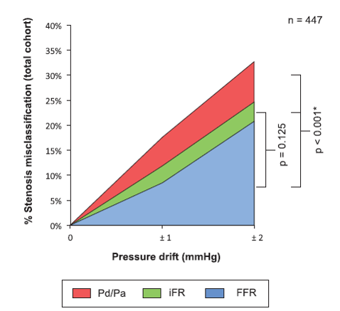
Compared to the hyperemic state, smaller separation between Pd and Pa at rest means even relatively small amounts of drift can lead to stenosis misclassification compared to FFR. That’s why it critical to detect even the slightest difference of pressure.
Use of FFR vs RFR16
| Uses of FFR | RFR | FFR |
|---|---|---|
| Left Main Coronary Artery22 | – | +++ |
| Systolic Dysfuntion23 | – | +++ |
| Post-PCI Side Branch Interrogation24 | – | +++ |
| Post-PCI Assessment25 | – | +++ |
| Equivalent Assessment | ||
| Multi-Vessel Disease26 | +++ | +++ |
| Diabetes27 | +++ | +++ |
| Uses of RFR | ||
| Tandem Lesions28 | +++ | + |
| Diffuse Lesions29 | +++ | + |
| Left Anterior Descending30 | +++ | ++ |
| Acute Coronary Syndrome31 | +++ | ++ |
Chart courtesy of Ziad Ali, MD.
PPG: Pullback Pressure Gradient
PPG is a predictive tool that quantifies focal coronary disease to enable precise PCI decision-making for better patient outcomes.32-36
Why Measure PPG?
Enhance Actionable Insights
- PPG, a novel epicardial index, objectively quantifies focal epicardial disease to identify patients that are most likely to benefit from PCI32,33
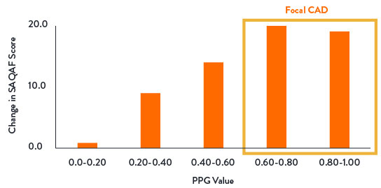
- PPG is a continuous metric; values approaching 1.0 represent focal CAD, whereas values close to 0 diffuse CAD33
- EAPCI has recognized and endorsed the value of PPG in Pre-procedural PCI Planning to identify patterns of focal and diffuse disease37

Predict Patient Benefit
By measuring PPG, you can predict which patients are most likely to be angina-free post-PCI.32,35
- PPG demonstrated excellent predictive capacity for optimal revascularization
- PPG was significantly correlated with the change in post-PCI FFR32
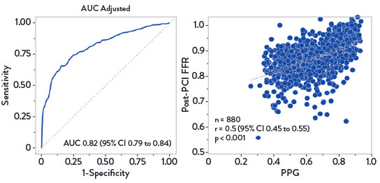
Predictive Capacity of PPG for FFR After PCI32
- Patients with high PPG (focal disease) reported greater improvement in angina and quality of life35
- PPG changed the revascularization decision in 1 out of 7 patients32
- Residual angina after PCI was ~2X more prevalent in patients with a low PPG (diffuse disease) than high PPG (focal)35
Change in Angina Frequency From Baseline to 1-Year Follow-Up36
- One year outcomes from the PPG Global Study showed:
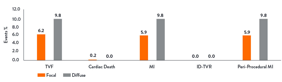
- PPG predicts suboptimal post-PCI physiology which is associated with higher rate of adverse events (TVF, MI, TVR).38
- PPG is the only independent predictor of angina improvement post-PCI within the study.38

TVF38
Safe and Simple Workflow
The PPG Global Study demonstrated that PCI was safer to perform in patients with focal disease, resulting in lower rates of periprocedural MI.36
PPG integrates seamlessly into epicardial assessment workflow as a part of Abbott's Full Physiology solution.3,32,39

*As compared to all commercially available full physiology solutions in the U.S. as of Q1, 2026. Refer to IFUs for additional information. Full physiology refers to: Fractional Flow Reserve (FFR), Resting Full-cycle Ratio (RFR), Index of Microcirculatory Resistance (IMR), Coronary Flow Reserve (CFR) and Pullback Pressure Gradient (PPG).
References:
- Pijls, NH., et al. Measurement of fractional flow reserve to assess the functional severity of coronary-artery stenoses. N Engl J Med. 1996;334(26):1703-1708.
- Volcano Corp. Verrata‡ guidewire and PrimeWire Prestige‡ Plus guidewire IFUs, Opsens Inc. OptoWire‡ guidewire and OptoWire‡ II guidewire IFUs, ACIST Medical Systems. Navvus‡ Microcatheter IFU, Boston Scientific Corporation. Comet‡ guidewire IFU. Refer to IFUs for additional information.
- PressureWire™ X Guidewire Instructions for Use (IFU). Refer to IFU for additional information.
- Pijls, NH., et al. Fractional Flow Reserve Versus Angiography for Guiding Percutaneous Coronary Intervention in Patients with Multivessel Coronary Artery Disease. 2-Year Follow-Up of the FAME Study. JACC. 2010; 56(3): 177-184.
- Fearon, WF., et al. Fractional Flow Reserve-Guided PCI as Compared with Coronary Bypass Surgery. N Engl J Med, Nov 4, 2021.
- Boutaleb AM, et al. Fractional flow reserve and non-hyperemic indices: Essential tools for percutaneous coronary interventions. World J Clin Cases. 2023;11(10):2123-2139.
- Johnson NP, et al. Continuum of Vasodilator Stress From Rest to Contrast Medium to Adenosine Hyperemia for Fractional Flow Reserve Assessment. JACC Cardiovasc Interv. 2016;9(8):757-767.
- Corcoran, D., et al. Fractional flow reserve: a clinical perspective. Int J Cardiovasc Imaging. 2017;33(7):961‐974.
- Pijls, NH., et al. Fractional flow reserve versus angiography for guiding percutaneous coronary intervention in patients with multivessel coronary artery disease: 2-year follow-up of the FAME (Fractional Flow Reserve Versus Angiography for Multivessel Evaluation) study. J Am Coll Cardiol. 2010;56(3):177‐184.
- Smits, PC., et al. Fractional flow reserve–guided multivessel angioplasty in myocardial infarction. N Engl J Med. 2017; 376:1234-1244.
- Svanerud, J., et al. Validation of a novel non-hyperaemic index of coronary artery stenosis severity: the Resting Full-cycle Ratio (VALIDATE RFR) study. EuroIntervention. 2018;14:806-814.
- Lee, JM., et al. Physiological and clinical assessment of resting physiological indexes. Circulation. 2019;139:889-900.
- Jeremias, A., et al. Resting full-cycle ratio (RFR): a novel physiologic index compared to Fractional Flow Reserve (FFR) in assessing the hemodynamic severity of a coronary stenosis: ILUMIEN I + PREDICT. EuroPCR 2018.
- Ahn, JM., et al. IRIS FFR: prognostic performance of five resting pressure-derived indexes of coronary physiology. TCT 2018.
- Nam, CW., et al. Fractional flow reserve assessment of serial lesions. Radcliffe Cardiology. April 2014.
- Ali, Z., Invasive Assessment of Coronary Physiology Is Here to Stay! TCT 2019.
- Kumar, G., et al. RE-VALIDATE: REal world VALIDATion of the non-hyperemic InDex of coronary Artery sTEnosis severity: resting full-cycle ratio (RFR) – RE-VALIDATE RFR. CRT 2019.
- Lee, JM., et al. Clinical Outcome of Lesions with discordant results among different invasive physiologic indices. Circulation Journal 2019 83:2210-2221.
- Jeremias, A., et al. A Test in Context: FFR: Accuracy, Prognostic Implications, and Limitations. J AM Coll Cardiol. 2017; 69:2748-58.
- Veer, M., et al. Comparison of Different Diastolic Resting Indexes to iFR. J AM Coll Cardiol. 2017;70(25):3088-3096.
- Cook, C. Quantification of the Effect of Pressure Wire Drift on the Diagnostic Performance of FFR, iFR, & Pd/Pa. Circ Cardiovasc Interv. 2016;9(4):e002988.
- Mallidi, J., et al. Long-term outcomes following fractional flow reserve-guided treatment of angiographically ambiguous left main coronary artery disease: a meta-analysis of prospective cohort studies. Catheter Cardiovasc Interv. 2015;86(1):12-18.
- Johnson, NP., et al. Diastolic pressure ratio: new approach and validation vs the instantaneous wave-free ratio. Eur Heart J. 2019; 40(31):2585-2594
- Koo, BK., et al. Physiologic assessment of jailed side branch lesions using fractional flow reserve. J Am Coll Cardiol. 2005;46(4):633-637.
- Johnson, NP., et al. Prognostic value of fractional flow reserve: linking physiologic severity to clinical outcomes. J Am Coll Cardiol. 2014;64(16):1641-1654.
- Escaned, J., et al. Clinical outcomes of state-of-the-art percutaneous coronary revascularization in patients with de novo three vessel disease: 1-year results of the SYNTAX II study. Eur Heart J. 2017;38(42):3124-3134.
- DEFINE-FLAIR Trial Investigators. Comparison of major adverse cardiac events between instantaneous wave-free ratio and fractional flow reserve–guided strategy in patients with or without type 2 diabetes. JAMA Cardiol. 2019;4(9):857-864.
- Nijjer, SS., et al. Pre-angioplasty instantaneous wave-free ratio pullback provides virtual intervention and predicts hemodynamic outcome for serial lesions and diffuse coronary artery disease. JACC Cardiovasc Interv. 2014;7(12):1386-96.
- Jeremias, A., et al. Blinded physiological assessment of residual ischemia after successful angiographic percutaneous coronary intervention: the DEFINE PCI study. JACC Cardiovasc Interv. 2019;12(20):1991-2001.
- Sen, S., et al. Clinical events after deferral of LAD revascularization following physiological coronary assessment. J Am Coll Cardiol. 2019;73(4):444-453.
- Escaned, J., et al. Safety of the deferral of coronary revascularization on the basis of instantaneous wave-free ratio and fractional flow reserve measurements in stable coronary artery disease and acute coronary syndromes. JACC Cardiovasc Interv. 2018;11(15):1437-1449.
- Collet, C., et al. Influence of Pathophysiologic Patterns of Coronary Artery Disease on Immediate Percutaneous Coronary Intervention Outcomes. Circulation. 2024:150:586-597.
- Collet, C., et al. Measurement of Hyperemic Pullback Pressure Gradients to Characterize Patterns of Coronary Atherosclerosis. J Am Coll Cardiol. 2019;74(14):1772–1784.
- Mizukami, T., et al. Procedural Outcomes After Percutaneous Coronary Interventions in Focal and Diffuse Coronary Artery Disease. J Am Coll Cardiol Intv. 2022:11(23).
- Collet, C., et al. Differential Improvement in Angina and Health-Related Quality of Life After Percutaneous Coronary Interventions in Focal and Diffuse Coronary Artery Disease. J Am Coll Cardiol Intv. 2022;15:2506-2518.
- Collet, C., Impact of Pullback Pressure Gradient (PPG) on Patient-Reported Outcomes in Patients with Coronary Artery Disease. Presented at TCT 2024.
- Escaned, J., et al. Applied coronary physiology for planning and guidance of percutaneous coronary interventions. A clinical consensus statement from European Association of Percutaneous Cardiovascular Interventions (EAPCI) of the European Society of Cardiology. EuroIntervention 2023; 19:464-481.
- Ikeda, K., et al. Impact of Pullback Pressure Gradient on Clinical Outcomes after Percutaneous Coronary Interventions. Circ Cardiovasc Interv. 2025:e016022.
- CoroFlow‡ Cardiovascular System Instructions for Use (IFU). Refer to IFU for additional information.
- Data on file at Abbott.
MAT–2512344 v2.0
Coroventis‡ CoroFlow‡ Cardiovascular System

Indications: CoroFlow‡ is indicated to provide hemodynamic information for use in the diagnosis of patients with cardiovascular diseases.
CoroFlow‡ is intended for use in catheterization and related cardiovascular specialty laboratories to compute and display various physiological parameters based on the output from one or more measuring devices.
Contraindications: The system has no patient alarm functions. Do not use for cardiac/vital signs monitoring.
Warnings:
- If CoroFlow‡ is used together with 3rd party infusion catheters for assessment of Absolute Flow and Resistance, ensure that the maximum infusion rate per manufacturers instruction is not exceeded or vessel injury may occur.
- Do not use the CoroFlow‡ Cardiovascular System if there is reason to believe the system's security has been compromised or if the system was unaccounted for a period of time (i.e. misappropriated, modified or tampered with).
- Do not leave the CoroFlow‡ Cardiovascular System unattended when logged in as a PC Administrator.
- To protect the privacy and security of sensitive information, including electronic protected health information (EPHI), and to protect the integrity of the system itself, the system should be located in a physically secure, access-controlled environment.
- To protect the privacy and security of sensitive information, including electronic protected health information (EPHI), the PC on to which CoroFlow‡ is installed must be configured according to the Installation Instructions in this manual. Failure to configure the PC correctly may result in increased risk for unauthorized release of protected health information. Windows settings include:
- Activation and configuration of restricted user Access
- Activation of Windows Firewall and blocking of network connections
- Activation of Windows Bitlocker drive encryption
- Activation of Windows Secure Boot
- Activation of Windows Anti-Virus scanning and ransomware protection. Ensure CoroFlow‡ is added in the list of trusted applications.
- Activation of Windows update
- Disable unused interfaces
- Use of this equipment adjacent to or stacked with other equipment should be avoided because it could result in improper operation. If such use is necessary, this equipment and the other equipment should be observed to verify that they are operating normally.
- Use of accessories, transducers and cables other than those specified or provided by Coroventis‡ could result in increased electromagnetic emissions or decreased electromagnetic immunity of this equipment and result in improper operation.
- Portable RF communications equipment (including peripherals such as antenna cables and external antennas) should be used no closer than 30 cm (12 inches) to any part of CoroFlow‡, including cables specified by Coroventis‡. Otherwise, degradation of the performance of this equipment could result.
Precautions:
- The PC and CoroHub‡ shall not be placed within the patient environment (1.5 m from patient).
- For operation of other devices used in conjunction with CoroFlow‡ consult the IFU for each of these devices for details on indication, handling and safety information.
- It is recommended to ensure local routines for data backup of stored recordings. CoroFlow‡ does not create backup of stored data.
- Always check minimum performance requirement on PC to ensure compatibility with CoroFlow‡.
- It is recommended to install CoroFlow‡ on a PC with backup battery to avoid interruption in case of power failure.
- Always manually review and confirm valid cursor positions and detected heart beats.
- Ensure that Pa and Pd pressure waveforms are aligned in phase and offset after equalization, or indices can be mis-calculated.
- Confirm that the correct Wi-Box is selected by manually matching the Wi-Box ID number with the Wi-Box in the lab.
- Changing parameter settings outside of default values may affect measurement performance, only for research purposes.
- Only to be used by healthcare professionals.
- Using a network location to store data may cause previously unidentified risks if the network malfunctions.
- No modification or tampering with CoroFlow‡ is permitted.
- CoroFlow‡, including accessories and components, shall not be used if it has been subject to damage.
- The assembly of medical electrical systems and modifications during the actual service life require evaluation to the requirements according to IEC 60601-1 standard series.
- CoroHub‡ does not have any serviceable parts and require no field maintenance. No modification or tampering with CoroHub‡ is permitted.
- CoroHub‡ shall not be immersed in liquid.
- CoroHub‡ shall not be used if it has been subject to damage.
- PPG values may be non-unique and different combinations or focal/diffuse disease may result in the same PPG value.
- Direct connection to a non-secure network, like the internet, may interfere with correct operation and/or result in inappropriate access to patient information. Furthermore, it should be noted that reconfiguring a used network may lead to inability to import patient as well as export examination data, ultimately leading to a risk of loss of patient and examination data. To avoid this problem Coroventis‡ recommends verifying network settings in the system setup after each change. The same caution is relevant regarding connection to DICOM.
- Always confirm valid pressure tracings, marker positions and selected beats.
- Resetting CoroHub‡ will reset PressureWire connections and Zeroing/Equalization parameters.
MAT-2007904 v4.0
PressureWire™ X Guidewire
INDICATIONS
The PressureWire™ X Guidewire is indicated to direct a catheter through a blood vessel and to measure physiological parameters in the heart and in the coronary and peripheral blood vessels. Physiological parameters include blood pressure. The PressureWire™ X Guidewire can also measure blood temperature.
CONTRAINDICATIONS
PressureWire™ X Guidewire is contraindicated for use in the cerebral vasculature.
POTENTIAL ADVERSE EVENTS
Potential adverse events which may be encountered during all catheterization procedures include, but are not limited to:
- Bleeding
- Hypotension / hypertension
- Chest pain
- Coronary / peripheral vascular injury
- Vascular dissection
- Perforation of vessels
- Stenosis
- Pseudoaneurysm
- Vasoconstriction
- Myocardial ischemia, tissue hypoxia
- Myocardial infarction
- Organ injury
- Thrombosis
- Death
- Arrhythmias
- Cardiac tamponade / pericardial effusion
- Bronchospasm (adenosine related)
- Prolonged procedure
- Foreign body in patient
- Urgent revascularization
- Congestive heart failure
- Embolus
- Local and / or systemic infection
- Device failures
- Hypersensitivity/ allergic reaction to device materials, medications or contrast
In addition, this device has a coating containing Polyethylene Glycol (PEG); potential allergic reactions (anaphylaxis) may occur during the interventional procedure if the patient is allergic to PEG.
WARNINGS
- The PressureWire™ X Guidewire must not be modified in any way by the customer. Making modifications may interfere with correct operation and will void product warranties. See your Abbott Technical Service representative for more information.
- Note the product “Use-by-date” on the package.
- The PressureWire™ X Guidewire is supplied sterile. Discard the guidewire if the pouch is opened or damaged, compromising the sterile barrier. The guidewire is designed for single use only and shall not be reused or resterilized. Adverse effects of using a non-sterile or resterilized guidewire may include, but are not limited to:
- Local and / or systemic infection
- Mechanical damage
- Inaccurate readings
- Observe all guidewire movements. Whenever the guidewire is moved or torqued, the tip movement should be examined under fluoroscopy. Never push, withdraw,
or torque the guidewire if it meets resistance or without observing corresponding movement of the tip; otherwise, vessel/ventricle trauma may occur. - Torquing or excessive manipulation of the guidewire in a sharp bend, against resistance, or repeated attempts to cross a total vessel occlusion may:
- Cause dissection or perforation of blood vessels
- Cause vessel spasm
- Damage and / or fracture the guidewire
- When introducing the guidewire, flush the catheter and administer anticoagulation as for a standard catheterization procedure or clotting may occur.
- Do not use the guidewire in the ventricles if the patient has a prosthetic mechanical or biological valve. It may result in damage to both the prosthesis and the guidewire, which may cause injury or death.
- Use of the PressureWire™ X Guidewire in conjunction with interventional devices with a short rapid exchange may result in a folded or fractured guidewire.
- High frequency surgical devices must not be used on a patient at the same time as the guidewire.
- The safety and effectiveness of the coated device have not been established, or are unknown, in vascular regions other than those specifically indicated.
- Failure to abide by the warnings in this Instructions for Use might result in damage to the device coating, which may necessitate intervention or result in serious adverse events.
PRECAUTIONS
- The PressureWire™ X Guidewire is a delicate instrument and should be handled carefully.
- Make sure that the PressureWire™ X Guidewire transmitter is kept dry to ensure accurate pressure and/or temperature readings. Inaccurate readings or a wet transmitter may necessitate device replacement.
- Do not use the PressureWire™ X Guidewire in conjunction with atherectomy catheters. It may damage the PressureWire™ X Guidewire.
- Do not withdraw or manipulate the PressureWire™ X Guidewire with a sharp-edged object. It may result in abrasion of the PressureWire™ X Guidewire coating. When wet, the hydrophilic coating increases the lubricity of the coated surface.
- Factors that may affect the accuracy of the diagnostic information include, but are not limited to:
- Improper placement of the aortic pressure sensor.
- Failure to achieve maximum coronary and myocardial hyperemia during FFR procedures
- Blood flow affected by the position of interventional devices, such as balloon catheters.
- PressureWire™ X Guidewire readings may be affected by defibrillation. Rezero the guidewire after defibrillation use.
- Do not measure pressure when the PressureWire™ X Guidewire sensor element is in a sharp bend or in contact with atrial or ventricular walls. It might result in pressure artifacts.
- Do not use the PressureWire™ X Guidewire together with another guidewire, for so-called jailed wire technique, due to difficulty in guidewire withdrawal and possible guidewire entrapment.
- Avoid abrasion of the distal hydrophilic and proximal PTFE coating regions. Use caution when manipulating, advancing and / or withdrawing these devices through needles, metal cannulas, stents, or other devices with sharp edges, or through tortuous or calcified blood vessels. Manipulation, advancement, and / or withdrawal past sharp or beveled edges may result in destruction and / or separation of the outer coating, which may lead to clinical adverse events, resulting in coating material remaining in the vasculature or device damage. This may result in adverse events requiring additional intervention.
- The integrity and performance of the device coating can be negatively impacted by preparation with incompatible media or solvents. Please take note of the following important recommendations:
- Avoid wiping the distal end of the device with dry gauze as this may damage the device coating; refer to Directions for Use for removal / reintroduction instructions.
- Avoid excessive wiping of the coated device.
- Avoid using alcohol, antiseptic solutions, or other solvents to pre-treat the device because this may cause unpredictable changes in the coating which could negatively affect the safety and performance of the device.
- Do not soak the device as it may adversely impact the hydrophilic coating on the device.
- The PressureWire™ X Guidewire contains parts with > 0.1% w/w concentration of 1-methyl-2-pyrrolidone or NMP, which has shown reproductive toxicity and teratogenicity in animal tests. This device has not been tested in pregnant women or in men intending to father children.
Effects on the developing fetus have not been studied. In the worst-case scenario, the amount of NMP that may elute out of the PressureWire™ X Guidewire is > 25 times lower than the permissible daily exposure (PDE) by the ICH Q3C (R8) Guidelines for Residual Solvents. Occurrence of this worst-case scenario is highly unlikely, and the expected exposure time of the patient for the FFR procedure is 60 minutes.
While there is no contraindication, the risks and reproductive effects of use of this device on humans are unknown at this time. It is recommended that the need of the patient and the clinical benefit of FFR be weighed against the risk stated above. - One or more components of this device may contain the following substances defined as CMR 1B in a concentration above 0.1% weight by weight: Cobalt; Chemical Abstracts Service (CAS) No. 7440-48-4; EC No. 231-158-0. Current scientific evidence supports that medical devices manufactured from stainless steel alloys containing cobalt do not cause an increased risk of cancer or adverse reproductive effects.
- The safety and effectiveness of the PressureWire™ X Guidewire has not been established in the pediatric population.
- Persons with known history of allergies to any of the components of this device listed below may suffer an allergic reaction to this device. Prior to its use on the patient, the patient should be counseled on the material contained in the device, and a thorough history of allergies must be discussed. This device contains:
- Epoxy adhesive
- Gold
- Palladium-iridium alloy
- Polyethylene glycol
- Polyimide
- Polyvinyl pyrrolidone hydrophilic coating
- Polytetrafluoroethylene coating
- Silicone
- Silicon
- Silicon nitride
- Stainless steel
- Tin solder paste
- Store at room temperature (15°C to 25°C) in a dry and dark place.
MAT-2103599 v4.0