Supera™ Stent

Results Matter. Design Matters.

  Supera Peripheral Stent System

Design Matters—Unique Interwoven Design of Supera™ Stent

Breakthrough Technology in Stent Platform

The Supera™ Stent’s unique design results in a platform with features unmatched among standard nitinol peripheral stents:


Strength—its high resistance to compression1

Strength


Low chronic outward force against vessel walls2

Low chronic outward force


High flexibility,3 which allows for fracture resistance4

High flexibility

View the Supera™ Stent Difference

The interwoven nitinol design provides high structural integrity and kink resistance, producing the features that set the Supera™ Stent apart.

Offering Unparalleled Strength for Compression Resistance

The Supera™ Stent exhibits more than 4 times the compression resistance of all other self-expanding nitinol stents, which are laser cut from an inflexible nitinol tube.2 Benchtop testing with the 5 pounds of force results in compression of a standard laser-cut nitinol stent (below left). When the same force is applied to the Supera™ Stent, it retains its circular form (below right).

In the clinical setting, those results are illustrated—by being deployed in the same patient and same vessel—via the angiogram and intravacular ultrasound (IVUS) images below. In the IVUS images, note the compression of the standard nitinol stent, while the Supera™ Stent was able to retain its circular shape. Such performance is particularly important in heavily calcified lesions.


  Laser-cut stent vs. Supera stent

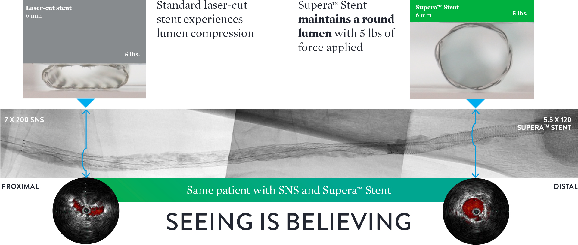
Source: Angiogram and IVUS images courtesy of Dr. Andrej Schmidt.

Exerting the Least Chronic Outward Force on the Vessel Wall1

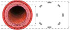
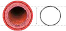
While laser-cut nitinol stents are designed and required to be oversized for deployment, the Supera™ Stent is unique in that it is sized 1:1 with the vessel diameter. So as seen in the image below, the laser-cut stents inherently exert force on the vessel wall throughout the lifetime of the stent, which in turn can result in endovascular injury over the long term. The lower chronic outward force (COF) with the Supera™ Stent results in fewer vessel injuries.1,5
 

Standard Nitinol Stent (SNS) Designs

Pushes against the vessel to open, through lifetime of SNS
Stent Oversizing High COF6Vessel Injury4
Chronic outward force (COF) is exerted on vessel by self-expanding stents due to inherent oversizing


Supera™ Stent

1:1 sizing allows to scaffold the vessel to maintain open
Sizing 1:1 Low COF6Minimal Vessel
Injury4
Supera™ Stent is sized 1:1 with the prepared vessel and therefore has minimal chronic outward force

Providing Unparalleled Flexibility3 and Zero Fractures4

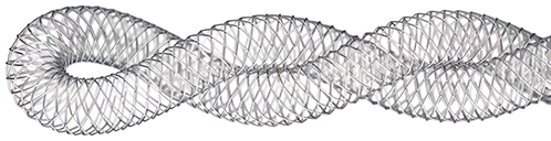
All other self-expanding nitinol stents are fashioned from a rigid, inflexible nitinol tube. As shown in the photo below, such laser-cut stents are less likely to conform to a dynamic vascular environment and can potentially kink and fracture in tortuous vessels (left).


Laser-Cut Stents

  Laser-cut stents

               Supera™ Stent

  Supera stent

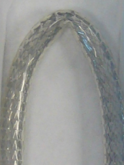
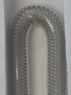
With the Supera™ Stent, however, individual flexible nitinol wires are interwoven for unparalleled flexibility,5 excellent kink resistance, and the ability to mimic the natural movement of the anatomy1,7,8 (right). Given the twisting and compression characteristics of the superficial femoral artery and proximal popliteal, this stent is an effective choice.

Consequently, data on over 2,000 patients published in 17 studies have shown that at 1 year the Supera™ Stent has zero fractures.4,9,10-24
 

0.6% Fractures

in 3 years across all studies4,9,10-24


Supera Stent
contact us

To learn more about Supera™ Stent, request a free demo and your local Abbott rep will be in touch shortly.

References

  1. Test(s) performed by and data on file at Abbott.
  2. Competitors tested include Astron Pulsar-18, Complete SE, EverFlex, Innova, LifeStent, Misago, S.M.A.R.T., and Zilver PTX. Test(s) performed by and data on file at Abbott.
  3. Flexibility is defined as kink resistance.  Competitors tested include Astron Pulsar-18, Complete SE,  EverFlex, Innova, LifeStent, Misago, S.M.A.R.T., and Zilver PTX. Test(s) performed by and data on file at Abbott.
  4. Garcia L. et al., Catheterization and Cardiovascular Interventions 2017 Jun 1;89(7):1259-1267
  5. Zhao HQ et al. Cardiovasc Intervent Radiol. 2009;32(4):720-6.
  6. Treitl, K.M., et al. European Radiology.2017; 10.1007.
  7. Arena F. et al., J Vasc Med Surg. 2013:1;116.
  8. Chen Y. et al., J Vasc Surg 2014;59:384-91.
  9. Brescia AA. et al., J Vasc Surg. 2015 Jun;61(6):1472-8
  10. Chan YC. et al., J Vasc Surg. 2015 Nov;62(5):1201-9.
  11. Dumantepe M. Vasc Endovascular Surg. 2017 Jul;51(5):240-246.
  12. George JC. et al., J Vasc Interv Radiol. 2014 Jun;25(6):954-61.
  13. Goltz JP. et al., J Endovasc Ther. 2012 Jun;19(3):450-6.
  14. León LR Jr. et al., J Vasc Surg. 2013 Apr;57(4):1014-22.
  15. Montero-Baker M. et al., J Vasc Surg. 2016 Oct;64(4):1002-8.
  16. Myint M. et al., J Endovasc Ther. 2016 Jun;23(3):433-41.
  17. Palena LM. et al., J Endovasc Ther. 2018 Oct;25(5):588-591.
  18. Scheinert D. et al., JACC Cardiovasc Interv. 2013 Jan;6(1):65-71.
  19. Scheinert D. et al., J Endovasc Ther. 2011 Dec;18(6):745-52.
  20. Steiner S. et al., J Endovasc Ther. 2016 Apr;23(2):347-55.
  21. Werner M. et al., EuroIntervention. 2014 Nov;10(7):861-8.
  22. San Norberto EM. et al., Ann Vasc Surg. 2017 May;41:186-195.
  23. Teymen B. et al., Vascular. 2018 Feb;26(1):54-61.
  24. Bhatt H. et al., Cardiovasc Revasc Med. 2018 Jul;19(5 Pt A):512-515.

MAT-2109824 v1.0

Important Safety Information
 

Supera™ Peripheral Stent System

rx-only-logo

Indications

The Supera™ Peripheral Stent System is indicated to improve luminal diameter in the treatment of patients with symptomatic de novo or restenotic native lesions or occlusions of the superficial femoral artery (SFA) and / or proximal popliteal artery with reference vessel diameters of 4.0 to 7.5 mm, and lesion lengths up to 140 mm.


Contraindications

The Supera™ Peripheral Stent System is contraindicated in:

  • Patients who are judged to have a lesion that prevents complete inflation of an angioplasty balloon or proper placement of the stent or stent delivery system.
  • Patients who cannot receive antiplatelet or anticoagulation therapy. Based on in vivo thrombogenicity testing, the device should not be used in patients who cannot be anticoagulated as there may be some thrombus formation in the absence of anticoagulation.


Warnings

  • This device is intended for single-use only. Do not reuse. Do not resterilize. Do not use if the package is opened or damaged.
  • Use this device prior to the “Use By” date as specified on the device package label. Store in a dry, dark, cool place.
  • DO NOT use if it is suspected that the sterility of the device has been compromised.
  • Persons with known hypersensitivities to Nitinol and / or its components (e.g. nickel-titanium) may suffer an allergic reaction to this implant.
  • Administer appropriate antiplatelet therapy pre- and post-procedure.
  • Careful attention should be paid when sizing and deploying the stent to prevent stent elongation. In the SUPERB clinical study, stent elongation was associated with a decrease in patency at 12 months.


Precautions

The Supera™ Peripheral Stent System should only be used by physicians and medical personnel trained in vascular interventional techniques and trained on the use of this device.

  • The long-term safety and effectiveness of the Supera™ Peripheral Stent System has not been established beyond three years.
  • The safety and effectiveness of the Supera™ Peripheral Stent System has not been established in patients who:
    • are less than 18 years old
    • are pregnant or lactating
    • have in-stent restenosis of the target lesion
    • have known hypersensitivity to any component of the stent system (e.g., nickel)
    • cannot tolerate contrast media and cannot be pre-treated
    • have uncontrolled hypercoagulability and / or another coagulopathy
  • This device is not designed for use with contrast media injection systems or power injection systems.
  • The flexible design of the Supera™ stent may result in variation in the deployed stent length.


Magnetic Resonance Imaging (MRI) Safety Information

Nonclinical testing has demonstrated that the Supera™ stent, in single and in overlapped configurations up to 250 mm in length, is MR Conditional. A patient with this device can be safely scanned in an MR system meeting the following conditions:

  • Static magnetic field of 1.5 or 3.0 Tesla
  • Maximum spatial gradient magnetic field of 2,500 Gauss/cm (25 T/m)
  • Maximum MR whole-body-averaged specific absorption rate (SAR) of
    • 2 W/kg for landmarks (i.e. center of RF coil) above the umbilicus
    • 1 W/kg for landmarks below the umbilicus and above the mid-thigh
    • 0.5 W/kg for landmarks below the mid-thigh

Under the scan conditions defined above, the Supera™ stent is expected to produce a maximum temperature rise of 7.6 °C after 15 minutes of continuous scanning.

In nonclinical testing, the image artifact caused by the device extends approximately 2 cm from the Supera™ stent when imaged with a gradient echo or spin echo sequence and a 3T MRI system.


Potential Adverse Events 

Potential adverse events include, but are not limited to:

  • Abrupt closure
  • Allergic reaction (contrast medium; drug; stent material)
  • Amputation or limb loss
  • Aneurysm or pseudoaneurysm in vessel or at vascular access site
  • Angina or coronary ischemia
  • Arrhythmia (including premature beats, bradycardia, atrial or ventricular tachycardia, atrial or ventricular fibrillation)
  • Arteriovenous fistula
  • Bleeding complications requiring transfusion or surgical intervention
  • Death
  • Detachment of a system component or implantation in an unintended site
  • Embolization, arterial or other (e.g. air, tissue, plaque, thrombotic material, or stent)
  • Emergent surgery
  • Fever
  • Hematoma or hemorrhagic event, with or without surgical repair
  • Hyperperfusion syndrome
  • Hypertension / Hypotension
  • Infection
  • Myocardial infarction
  • Pain (leg, foot, and/or insertion site)
  • Partial stent deployment
  • Peripheral nerve injury
  • Pulmonary embolism
  • Renal failure or insufficiency
  • Restenosis of vessel in stented segment
  • Shock
  • Stent malapposition or migration, which may require emergency surgery to remove stent
  • Stent strut fracture
  • Thrombosis or occlusion
  • Stroke
  • Transient ischemic attack
  • Venous thromboembolism
  • Vessel dissection, perforation or rupture
  • Vessel spasm or recoil
  • Worsening claudication or rest pain

MAT-2103597 v3.0