OPTIS™ Imaging Systems

OCT Imaging and Coronary Physiology on One Platform

OPTIS™ Imaging Systems

OPTIS™ Next Imaging Systems

OPTIS™ Integrated Next Imaging System and OPTIS™ Mobile Next Imaging System are the newest generation of Abbott’s imaging systems that are available with Ultreon 1.0 Software. Streamlined and intuitive, Ultreon™ 1.0 Software gives better insights to optimize patient outcomes through automation and an improved workflow.1-4 The OPTIS™ Next Systems use high-powered processors that support AI technology for faster information display and workflow efficiency.1

OPTIS™ Integrated Next Imaging System: It's always on, it's always ready

OPTIS™ Integrated Next System is installed in a single cath lab; it's always on and always ready to perform intravascular imaging and coronary physiology.

OPTIS™ Integrated Next Imaging System set up
OPTIS™ Integrated Next Imaging System set up. Components shown include Wi-Box™ AO Transmitter, DOC, TSC, and OCT user interface display on the Boom Monitor.

OPTIS™ Integrated Next System provides benefits to the physicians:

  • Save setup time and start the PCI procedure right away because the system is always on and ready to use
  • Provides the user with a wireless tableside control (TSC) of OCT and RFR/FFR acquisition and review at a procedure table. The TSC also allows for single-operator control of the procedure if needed.
  • Streamlines the PCI procedure by having OCT and coronary physiology always available to guide PCI decision-making
  • Compatible with Dragonfly OpStar™ Imaging Catheter, Dragonfly™ OPTIS Imaging Catheter
     

Consideration for use:

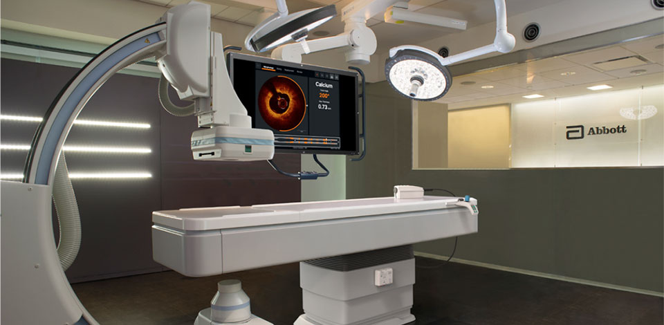
The OPTIS™ Integrated Next System is the latest integrated imaging system that supports Ultreon™ 1.0 Software. It is recommended for imaging and physiology needs, for busy cath labs and high PCI volume centers when the availability of OCT and/or coronary physiology is always needed without advanced arrangements.

image
OPTIS™ Tableside Controller Next (TSC)

OPTIS™ Mobile Next Imaging System: flexibility for multiple cath labs

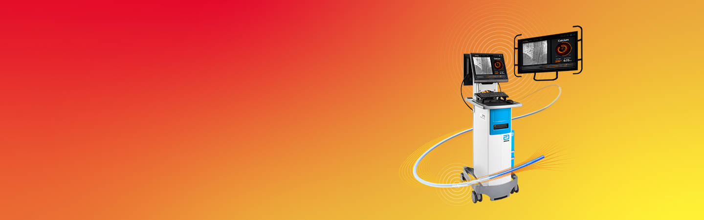
OPTIS™ Mobile Next Imaging System is a transportable system designed for use in multiple cath labs with easy pre-installed connections.

OPTIS™ Mobile Next Imaging System set up with an added QUANTIEN™ Measurement System on the table side. Components shown include Wi-Box™ AO Transmitter, DOC, TSC, and OCT user interface display on the Boom monitor

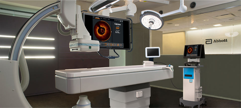
The OPTIS™ Mobile Next System provides the same functionality as the OPTIS™ Integrated Next System, but provides mobile system workflow via an easily transportable cart to serve multiple cath labs.

  • Integrates into the cath lab via easy pre-installed connections
  • Accommodates staggered PCI procedures at different times or on different days
  • One transportable cart to support PCI in multiple cath labs
  • Compatible with Dragonfly OpStar™ Imaging Catheter, Dragonfly OPTIS™ Imaging Catheter
     

Consideration for use:

The OPTIS™ Mobile Next Imaging System is the latest mobile imaging system that supports Ultreon™ 1.0 Software. It is recommended for multiple cath labs that require flexibility or for busy cath labs that already have an OPTIS™ Integrated Next Imaging System and need an additional system as a back up.
 

References

OPTIS™ Next Imaging Systems IFU. Refer to Instructions For Use (IFU) for additional information.

  1. Data on File at Abbott.
  2. Zhang J, et al. Intravascular ultrasound versus angiography-guided drug-eluting stent implantation: the ULTIMATE trial. J Am Coll Cardiol. 2018;72(24):3126-3137.
  3. Hong M et al., IVUS-XPL 5 Year Outcomes, TCT 2019.
  4. Jones et al. JACC Cardiovascular Interventions, 2018, vol 11 (14). “Angiography Alone Versus Angiography Plus Optical Coherence Tomography to Guide Percutaneous Coronary Intervention – Outcomes From the Pan-London PCI Cohort”.

MAT-2106765 v1.0

Important Safety Information
 

OPTIS™ Next Imaging Systems and Software

Indications

The Ultreon™ 1.0 Software is intended to be used only with compatible OPTIS™ Next Imaging Systems. The OPTIS™ Next Imaging System with a compatible Dragonfly™ OPTIS™ Imaging Catheter or Dragonfly OpStar™ Imaging Catheter is intended for the imaging of coronary arteries and is indicated in patients who are candidates for transluminal interventional procedures. The Dragonfly™ OPTIS™ Imaging Catheter or Dragonfly OpStar™ Imaging Catheter is intended for use in vessels 2.0 to 3.5 mm in diameter. The Dragonfly™ OPTIS™ Imaging Catheter or Dragonfly OpStar™ Imaging Catheter is not intended for use in the left main coronary artery or in a target vessel which has undergone a previous bypass procedure. The OPTIS™ Next Imaging System is intended for use in the catheterization and related cardiovascular specialty laboratories and will further compute and display various physiological parameters based on the output from one or more electrodes, transducers, or measuring devices. The physician may use the acquired physiological parameters, along with knowledge of patient history, medical expertise, and clinical judgment to determine if therapeutic intervention is indicated.

Contraindications: Use of the Ultreon™ 1.0 Software is contraindicated where introduction of any catheter would constitute a threat to patient safety.
Contraindications include:

  • Bacteremia or sepsis
  • Major coagulation system abnormalities
  • Patients diagnosed with coronary artery spasm
  • Patients disqualified for coronary artery bypass graft (CABG) surgery
  • Patients disqualified for percutaneous transluminal coronary angioplasty (PTCA)
  • Severe hemodynamic instability or shock
  • Total occlusion
  • Large thrombus
  • Acute renal failure
  • Inability to tolerate systemic anticoagulation is a contraindication to use of OCT for coronary imaging.
  • PressureWire™ Guidewire is contraindicated for use in the cerebral vasculature.
  • The system has no patient alarm functions. Do not use for cardiac monitoring.


Complications: The risks involved in vascular imaging include those associated with all catheterization procedures. The following complications may occur as a consequence of intravascular imaging and may necessitate additional medical treatment including surgical intervention.

  • Abnormal heart rhythm or arrhythmias
  • Acute myocardial infarction
  • Allergic reaction to the contrast media or drug administered for the procedure
  • Arterial dissection, injury, or perforation
  • Bleeding
  • Catheter access site reactions: sterile inflammation or granuloma
  • Coronary artery spasm
  • Death
  • Embolism
  • Myocardial ischemia
  • Renal insufficiency or failure from contrast media use
  • Repeat revascularization
  • Thrombus formation, abrupt closure, or total occlusion
  • Tissue necrosis
  • Unstable angina
  • Hypotension
     

Warnings:

  • Refer to the contrast media Instructions for Use for general warnings and precautions relating to use of contrast media.
  • The heart rate and mean pressure values shown on the OPTIS™ Next Imaging System are for reference only and are not intended to be used as the primary display.
  • The system may place the point of index value at the wrong location due to pressure artifacts, for example: abnormal heartbeats, artifacts in AO (Pa) caused by flushing of guiding catheter, or valve opening / closing. The physician should always confirm that the point selected by the system is a valid point for the calculation of index value.
  • Inside the catheterization laboratory, only port-powered USB drives may be connected to the USB port. Connecting externally powered devices to the USB port in the patient vicinity may compromise electrical isolation and cause patient injury.
  • To protect the privacy and security of sensitive information, including electronic protected health information (EPHI), and to protect the integrity of the system itself, the system should be located in a physically secure, access-controlled environment. Do not use the OPTIS™ Next Imaging System if there is reason to believe the system’s security has been compromised or if the system was unaccounted for during a period of time (i.e., misappropriated, modified, or tampered with).
     

Precautions:

  • Safety and effectiveness have been established for the following patient population: adult patients undergoing non-emergent percutaneous coronary interventions in lesions with reference vessel diameters between 2.0 to 3.5 mm, which were not located in the left main coronary artery or in a target vessel which has undergone previous bypass procedures.
  • Monitor the OCT image for indications of Dragonfly™ Imaging Catheter optical failure. If optical failure is suspected, remove the Dragonfly™ Imaging Catheter from the patient, press “Unload” on the drive motor and optical controller (DOC), detach the catheter, and replace it with a new one.
  • If the pullback triggers before contrast is injected, repeat the pullback.
  • For optimal imaging, only use 100% contrast media.

MAT-2104193 v3.0