OPTIS™ Imaging Systems

OCT Imaging and Coronary Physiology on One Platform

HeaderBanner

Cath Lab System Integration with OPTIS™ Imaging Systems

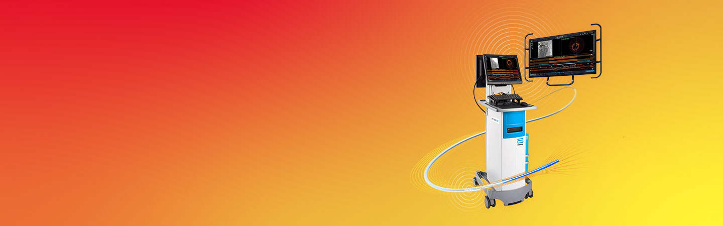
Abbott’s OPTIS™ Imaging Systems are easily integrated with the cath lab systems.
 

Integration of FFR/RFR* with cath lab recording hemodynamic system via Wi‑Box™ AO Transmitter connection

The OPTIS™ Imaging System displays coronary physiology (FFR/RFR*) measurement using a wireless PressureWire™ X Guidewire and an external aortic pressure (AO) transducer. Aortic pressure signal is sent from an AO transducer to Wi-Box™ AO Transmitter and wirelessly transmitted to the OPTIS™ Imaging System.1

Wi-Box™ AO Transmitter sends signal wireless to the OPTIS™ Integrated System. Components shown include Wi-Box™ AO Transmitter, DOC, TSC and OCT user interface display on the Boom Monitor

Wi-Box™ AO Transmitter is a small device easily installed by mounting on the base of the cath lab table. No cables to connect to OPTIS™ Imaging System, no clutter, no interference with the flow of the PCI procedure. Wi-Box™ AO Transmitter sends the AO pressure signal wirelessly to the OPTIS™ Imaging System and the output is displayed on the existing cath lab monitors.

DICOM Integration

OPTIS™ Imaging Systems implement several DICOM services for exchanging information and images. These systems allow for the querying of patient information from a network storage device, as well as the storing of acquired OCT images and physiology images, as “secondary capture” images, to the network storage device or CD/DVD.
 

DICOM integration features:2

  • DICOM Modality Worklist Query (results shown in Add Patient wizard)
  • Export to DICOM storage server
  • Export to USB (DICOM)
  • Export to USB (raw)

To view DICOM conformance statement, please click here.

DICOM=Digital Imaging and Communication in Medicine

Connection to the Boom monitor

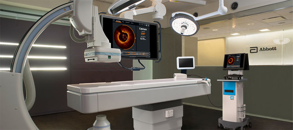
OPTIS™ Imaging Systems can be easily connected to standard Boom monitors in the cath lab to display the OCT user interface on the boom monitor to view PCI information from the procedure table.

OPTIS™ Integrated System
OPTIS™ Integrated System, OPTIS™ Mobile System and an added QUANTIEN™ Measurement System set up. Components shown include Wi-Box™ AO Transmitter, DOC, TSC and OCT user interface displayed on the Boom Monitor.

Connection options:

  1. OPTIS™ Integrated Imaging System is connected to Boom Monitor via DVI Extender
  2. OPTIS™ Mobile System is connected via Connectivity Boom Box

For more details on the DVI extender and Connectivity Box, see system components in the OPTIS™ Imaging Systems page.

Connection to cath lab angio systems

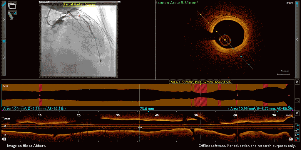
The OPTIS ™ Imaging Systems can be integrated with the cath lab angio systems to display OCT and angio co-registration (ACR) on the same screen.

img10_208

OCT and angio co-registration (ACR)

*RFR is available with AptiVue™ Imaging Software and Ultreon™ 1.0 Software.
 

Notes

  1. Compatible with most of hemodynamic systems in the cath lab that are following ANSI/AAMI standards.
  2. Available with AptiVue™ Imaging Software.
     

References

Wi-Box™ AO Transmitter and OPTIS™ Imaging Systems and Software IFUs. Refer to Instructions For Use (IFU) for additional information.

MAT-2106761 v1.0

Important Safety Information

OPTIS™ Imaging Systems and Software

rx-only-logo

Indications The OPTIS™ Software and AptiVue™ E Series Software are intended to be used only with compatible OPTIS™ Imaging Systems.

The OPTIS™ Imaging System with a compatible Dragonfly™ Imaging Catheter is intended for the imaging of coronary arteries and is indicated in patients who are candidates for transluminal interventional procedures. The compatible Dragonfly™ Imaging Catheters are intended for use in vessels 2.0 to 3.5 mm in diameter. The compatible Dragonfly™ Imaging Catheters are not intended for use in the left main coronary artery or in a target vessel which has undergone a previous bypass procedure.

The OPTIS™ Imaging System is intended for use in the catheterization and related cardiovascular specialty laboratories and will further compute and display various physiological parameters based on the output from one or more electrodes, transducers, or measuring devices. The physician may use the acquired physiological parameters, along with knowledge of patient history, medical expertise and clinical judgment to determine if therapeutic intervention is indicated.

Contraindications: The OPTIS™ Integrated System and Mobile System with Software are contraindicated where introduction of any catheter would constitute a threat to patient safety. Contraindications include:

  • Bacteremia or sepsis
  • Major coagulation system abnormalities
  • Patients diagnosed with coronary artery spasm
  • Patients disqualified for CABG surgery
  • Patients disqualified for PTCA
  • Severe hemodynamic instability or shock
  • Total occlusion
  • Large thrombus
  • Acute renal failure

NOTE: The systems have no patient alarm functions. Do not use for cardiac monitoring.
 

Warnings:

  • Appropriate anticoagulant and vasodilator therapy must be used during the procedure as needed.
  • Observe all advancement and movement of the Dragonfly™ Imaging Catheter under fluoroscopy. Always advance and withdraw the catheter slowly. Failure to observe device movement fluoroscopically may result in vessel injury or device damage.
  • Leave the guidewire engaged with the catheter at all times during use. Do not withdraw or advance the guidewire prior to withdrawing the catheter.
  • If resistance is encountered during advancement or withdrawal of the Dragonfly™ Imaging Catheter, stop manipulation and evaluate under fluoroscopy. If the cause of resistance cannot be determined or mitigated, carefully remove the catheter and guidewire together.
  • The Dragonfly™ Imaging Catheter should never be forced into lumens that are narrower than the catheter body or forced through a tight or heavily calcified lesion.
  • The Dragonfly™ Imaging Catheter should not be advanced through abnormally tortuous anatomy.
  • When advancing or retracting a catheter with a monorail tip through a stented vessel, the catheter may engage the stent between the junction of the Dragonfly™ Imaging Catheter and guidewire, resulting in entrapment of catheter/guidewire, catheter tip separation, and/or stent dislocation.
  • Refer to the contrast media’s instructions-for-use for general warnings and precautions relating to use of the contrast media.
  • To protect the privacy and security of sensitive information, including electronic protected health information (EPHI), and to protect the integrity of the system itself, the system should be located in a physically secure, access controlled environment.
  • Do not use the OPTIS™ Imaging System if there is reason to believe the system's security has been compromised or if the system was unaccounted for a period of time (i.e. misappropriated, modified or tampered with).


Precautions:

  • Safety and effectiveness have been established for the following patient population: adult patients undergoing non-emergent percutaneous coronary interventions in lesions with reference vessel diameters between 2.0 to 3.5 mm, which were not located in the left main coronary artery or in a target vessel which has undergone previous bypass procedures.
  • For optimal imaging, only use 100% contrast media.
  • Store the Dragonfly™ Imaging Catheter at ambient temperature in a dry location out of direct sunlight.
  • Never attempt to attach or detach a Dragonfly™ Imaging Catheter to the DOC while the “lock” LED is lit.
  • Do not kink, sharply bend, pinch, or crush the Dragonfly™ Imaging Catheter at any time.
  • The Dragonfly™ Imaging Catheter is for single use only. Do not reuse, re-sterilize, or reprocess.
  • The Dragonfly™ Imaging Catheter is sterilized by ethylene oxide and is intended for one time use only. Non-pyrogenic. Do not use if the package is opened or damaged.
  • After use, the Dragonfly™ Imaging Catheter may be a potential biohazard. Handle and dispose of in accordance with accepted medical practice and applicable laws and regulations.
  • The Dragonfly™ Imaging Catheter has no user serviceable parts. Do not attempt to repair or alter any part of the catheter assembly as provided.

MAT-2115909 v2.0

OPTIS™ Next Imaging Systems and Software

Indications

The Ultreon™ 1.0 Software is intended to be used only with compatible OPTIS™ Next Imaging Systems. The OPTIS™ Next Imaging System with a compatible Dragonfly™ OPTIS™ Imaging Catheter or Dragonfly OpStar™ Imaging Catheter is intended for the imaging of coronary arteries and is indicated in patients who are candidates for transluminal interventional procedures. The Dragonfly™ OPTIS™ Imaging Catheter or Dragonfly OpStar™ Imaging Catheter is intended for use in vessels 2.0 to 3.5 mm in diameter. The Dragonfly™ OPTIS™ Imaging Catheter or Dragonfly OpStar™ Imaging Catheter is not intended for use in the left main coronary artery or in a target vessel which has undergone a previous bypass procedure. The OPTIS™ Next Imaging System is intended for use in the catheterization and related cardiovascular specialty laboratories and will further compute and display various physiological parameters based on the output from one or more electrodes, transducers, or measuring devices. The physician may use the acquired physiological parameters, along with knowledge of patient history, medical expertise, and clinical judgment to determine if therapeutic intervention is indicated.

Contraindications: Use of the Ultreon™ 1.0 Software is contraindicated where introduction of any catheter would constitute a threat to patient safety.
Contraindications include:

  • Bacteremia or sepsis
  • Major coagulation system abnormalities
  • Patients diagnosed with coronary artery spasm
  • Patients disqualified for coronary artery bypass graft (CABG) surgery
  • Patients disqualified for percutaneous transluminal coronary angioplasty (PTCA)
  • Severe hemodynamic instability or shock
  • Total occlusion
  • Large thrombus
  • Acute renal failure
  • Inability to tolerate systemic anticoagulation is a contraindication to use of OCT for coronary imaging.
  • PressureWire™ Guidewire is contraindicated for use in the cerebral vasculature.
  • The system has no patient alarm functions. Do not use for cardiac monitoring.


Complications: The risks involved in vascular imaging include those associated with all catheterization procedures. The following complications may occur as a consequence of intravascular imaging and may necessitate additional medical treatment including surgical intervention.

  • Abnormal heart rhythm or arrhythmias
  • Acute myocardial infarction
  • Allergic reaction to the contrast media or drug administered for the procedure
  • Arterial dissection, injury, or perforation
  • Bleeding
  • Catheter access site reactions: sterile inflammation or granuloma
  • Coronary artery spasm
  • Death
  • Embolism
  • Myocardial ischemia
  • Renal insufficiency or failure from contrast media use
  • Repeat revascularization
  • Thrombus formation, abrupt closure, or total occlusion
  • Tissue necrosis
  • Unstable angina
  • Hypotension
     

Warnings:

  • Refer to the contrast media Instructions for Use for general warnings and precautions relating to use of contrast media.
  • The heart rate and mean pressure values shown on the OPTIS™ Next Imaging System are for reference only and are not intended to be used as the primary display.
  • The system may place the point of index value at the wrong location due to pressure artifacts, for example: abnormal heartbeats, artifacts in AO (Pa) caused by flushing of guiding catheter, or valve opening / closing. The physician should always confirm that the point selected by the system is a valid point for the calculation of index value.
  • Inside the catheterization laboratory, only port-powered USB drives may be connected to the USB port. Connecting externally powered devices to the USB port in the patient vicinity may compromise electrical isolation and cause patient injury.
  • To protect the privacy and security of sensitive information, including electronic protected health information (EPHI), and to protect the integrity of the system itself, the system should be located in a physically secure, access-controlled environment. Do not use the OPTIS™ Next Imaging System if there is reason to believe the system’s security has been compromised or if the system was unaccounted for during a period of time (i.e., misappropriated, modified, or tampered with).
     

Precautions:

  • Safety and effectiveness have been established for the following patient population: adult patients undergoing non-emergent percutaneous coronary interventions in lesions with reference vessel diameters between 2.0 to 3.5 mm, which were not located in the left main coronary artery or in a target vessel which has undergone previous bypass procedures.
  • Monitor the OCT image for indications of Dragonfly™ Imaging Catheter optical failure. If optical failure is suspected, remove the Dragonfly™ Imaging Catheter from the patient, press “Unload” on the drive motor and optical controller (DOC), detach the catheter, and replace it with a new one.
  • If the pullback triggers before contrast is injected, repeat the pullback.
  • For optimal imaging, only use 100% contrast media.

MAT-2104193 v3.0